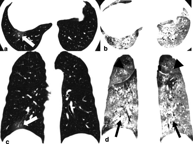
Fig. 2.
A 61-year-old male with COVID-19 in the death group. a, c Transverse and coronal CT images with score of 2 (1 [1–25%] * 2 [GGO]) showed GGO (white arrow) in the right lower lobe when the patient underwent CT scan on the same day with fever onsets. b, d Transverse and coronal CT images with score of 40 (calculated as 3 [50–75% distribution in right upper lobe] * 2 [GGO, black arrow head] + 3 [50–75% distribution in left upper lobe] * 2 [GGO, black arrow head] + 4 [> 75% distribution in right lower lobe] * 3 [consolidation, black arrow] + 4 [> 75% distribution in left lower lobe] * 3 [consolidation, black arrow] + 2 [GGO, not showed] * 2 [25–50% distribution in right middle lobe]) showed lesions in whole lung lobes 9 days after onset (10 days before death)

