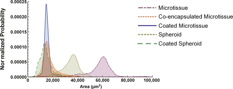
Figure 7.
Projected surface area distributions of 3D human liver models. Collagen-based microtissues containing encapsulated PHHs were fabricated using the droplet microfluidic device shown in Figure 1, while self-assembled spheroids were created as shown in Figure 3. Collagen-based microtissues containing encapsulated PHHs and 3T3-J2 fibroblasts were fabricated to create coencapsulated microtissue. 3T3-J2 fibroblasts were coated onto the PHH microtissues or spheroids to create coated microtissue and coated spheroid, respectively. Spheroid and Microtissue indicate models with only PHHs. After 21 days of culture, 375 individual constructs for each model were fixed and analyzed via phase-contrast microscopy for projected surface area determinations. Shown here are the histograms of the projected surface areas for the five culture/coculture models.

