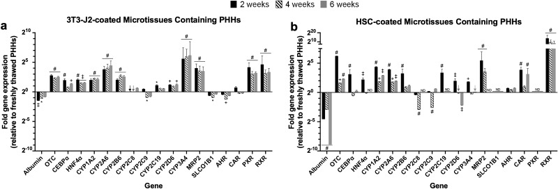
Figure 9.
Long-term gene expression in 3T3-J2-coated and HSC-coated microtissues. 3T3-J2 fibroblasts or primary human HSCs were coated onto the surface of the polymerized collagen-based PHH microtissues and cultured for ∼6 weeks. Quantitative gene expression of albumin, ornithine transcarbamylase (OTC), hepatic transcription factors (CEBPa and HNF4a), CYP enzymes (CYP1A2, CYP2A6, CYP2B6, CYP2C8, CYP2C9, CYP2C19, CYP2D6, and CYP3A4), canalicular transporter (MRP2), basolateral transporter (SLCO1B1), and nuclear receptors (AHR, CAR, PXR, and RXR) in 3T3-J2-coated microtissues (a) and HSC-coated microtissues (b) at 2, 4, and 6 weeks as compared to freshly thawed PHHs (line 20) that were immediately lysed in suspension for RNA (i.e., expression levels in the coated microtissues at the 20 line are near identical to the levels in freshly thawed PHHs). Arrows indicate detectable levels at the 20 line. For expression levels denoted ND (not detectable), amplification was not observed for the given gene within 40 cycles of quantitative polymerase chain reaction (qPCR). Statistical significance for gene expression levels is displayed for 3T3-J2 coated and HSC coated at 2, 4, and 6 weeks relative to freshly thawed PHHs (*p ≤ 0.05, +p ≤ 0.01, ‡p ≤ 0.001, #p ≤ 0.0001).

