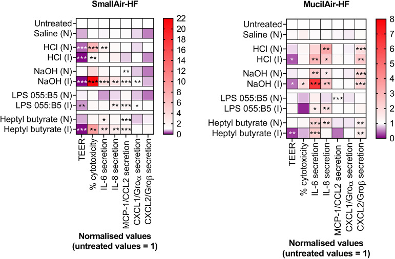
Figure 2.
Changes in markers of cellular- and tissue-specific KEs in SmallAir-HF™ (left) and MucilAir-HF™ (right) models following a single-dose exposure by aerosol (N) or by direct inoculation (I). Cellular-specific KE included percentage (%) cytotoxicity, cytokines (IL-6, IL-8) and chemokines (MCP-1/CCL2, CXCL1/Groα, CXCL2/Groβ) secretion. Tissue-specific KE included epithelial barrier impairment, quantified as changes in TEER. Data are presented as mean and normalized to untreated cultures. Symbols (*), (**), and (***) indicate p < 0.05, p < 0.01, and p < 0.001, respectively (two-way ANOVA followed by Dunnett post-test; comparison to the untreated controls).

