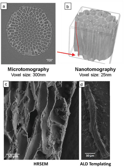
Figure 4.

a) Cross section of Jania sp. imaged at ID19 of the ESRF using X‐ray microtomography. b) 3D reconstruction of Jania sp. based on high‐resolution X‐ray nanotomography performed at ID16B of the ESRF showing the helical microstructure discovered in Jania sp. The red dashed line and red arrow highlight a spiraling pore edge. Sample height is 54 µm. c) Longitudinal cross section of Jania sp. imaged by HRSEM. A helix is observed in the cross section. d) Alumina replica of one of the Jania sp. inner pores obtained via ALD process, imaged by HRSEM.
