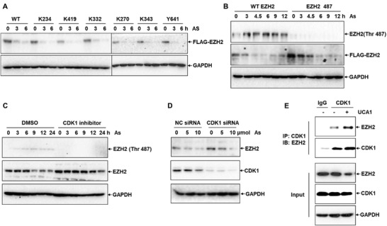
Figure 4.

LncRNA UCA1 promotes the degradation of EZH2 via CDK1. A) The degradation of EZH2 in HepG2 cells transfected with FLAG‐EZH2 plasmids including mutated phosphorylation sites (K234, K419, K332, K270, K343, and Y641) in response to 10 µmol As for 0, 3, and 6 h was detected via Western blot analysis (n = 3). B) Western blot analysis of the phosphorylation of EZH2 at Thr‐487 site in HepG2 cells exposed to 10 µmol As for 0, 3, 6, 9, 12, and 24 h (n = 3). C) The phosphorylation of EZH2 and the protein level of EZH2 in HepG2 cells pretreated with 1 µmol CDK1 inhibitor responding to 10 µmol As at different time points (n = 3). D) Western blot analysis of the protein contents of EZH2 and CDK1 in NC siRNA control and CDK1 siRNA cells under indicated dosage of As (n = 3). E) Enrichment of EZH2 in HepG2 cells transfected with CDK1 and UCA1 plasmids from CDK1 antibody and normal IgG pulldown complexes was detected by Western blot assay in IP assay (n = 3).
