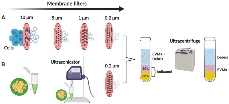
Figure 2.
Schematic diagram of generating EVMs. (A) Cells in suspension are extruded through a polycarbonate membrane using a mini-extruder, and (B) cells in suspension are ultrasonicated for 1 min. Crude EVMs are filtered through a 0.2 µm filter to produce EMVs smaller than 200 nm. EMVs are purified by two-step OptiPrep density gradient ultracentrifugation. EVMs: extracellular vesicle mimetics. Figure created with BioRender.

