Figure 1.
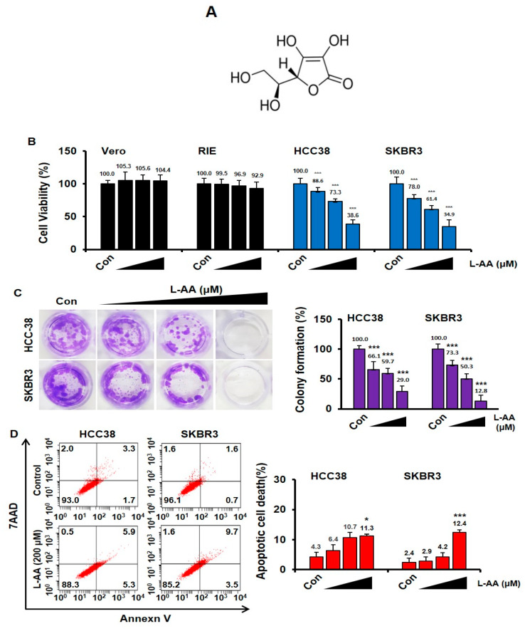
L-ascorbic acid (L-AA) inhibits breast cancer growth. (A) The chemical structure of L-AA. (B) Normal cells (Vero and rat intestinal epithelium (RIE)) and breast cancer cells (HCC38 and SKBR3) were treated with various concentrations of L-AA (50, 100, and 200 μM) for 48 h and then stained with trypan blue. Viable and dead cells were counted. (C) Breast cancer cells were treated with L-AA (50, 100, and 200 μM) every 3 days. After the 14 day treatment, cell colonies were stained with crystal violet. Crystal violet stained cells were extracted using 33% acetic acid and quantified by measuring absorbance at 570 nm on the microplate reader. (D) Breast cancer cells were treated with L-AA (50, 100, and 200 μM) for 48 h and then stained with annexin V-FITC and 7AAD in the binding buffer at room temperature in the dark. Stained cells were detected by LSRFortessa flow cytometry. The graph shows the sum of annexin V-FITC alone-positive cells (early apoptotic cells) and annexin V-FITC and 7AAD double positive cells (late apoptotic cells) from whole stained cells. Data are shown as the mean of three independent experiments, and the error bars represent standard deviation (SD). * p < 0.05 and *** p < 0.001 versus the control group.

