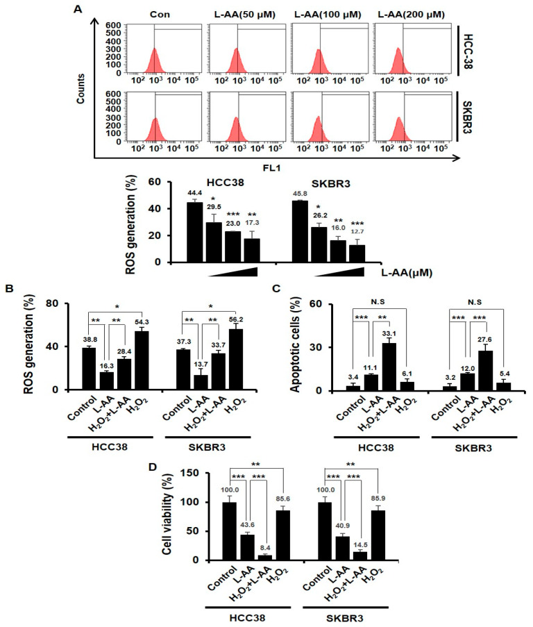
Figure 2.
L-AA-induced apoptosis is not correlated to the intracellular reactive oxygen species (ROS) generation. (A) Breast cancer cells (HCC38 and SKBR3) were treated with various concentrations of L-AA (50, 100, and 200 μM) for 24 h and then treated with H2DCF-DA fluorescent dye for 1 h at 37 °C. ROS generation was measured by LSRFortessa flow cytometry. Graph shows the proportion of H2DCF-DA-positive cells in the total cells. (B) Breast cancer cells were pre-treated with L-AA (200 μM) for 1 h, followed by exposure to H2O2 (10 μM) for 24 h. ROS production was measured. (C,D) HCC38 and SKBR3 cells were pre-treated with L-AA (200 μM) followed by exposure to H2O2 (10 μM) for 48 h. (C) Stained with annexin V-FITC and 7AAD and analyzed by LSRFortessa flow cytometry. The graph shows the proportion of annexin V-FITC alone-positive cells (early apoptotic cells) and annexin V-FITC and 7AAD double positive cells (late apoptotic cells) in whole stained cells. (D) Trypan blue assay was performed. Data are shown as the mean of three independent experiments, and the error bars represent standard deviation (SD). * p < 0.05, ** p < 0.01, and *** p < 0.001 were considered statistically significant.

