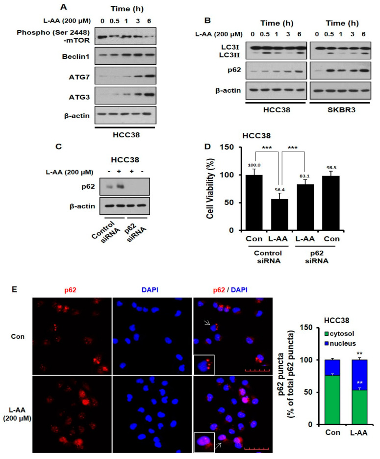
Figure 3.
L-AA induces autophagosome formation, while L-AA increases p62 accumulation in the nucleus. (A,B) Breast cancer cells were treated with L-AA (200 μM) for indicated hours (0–6 h) and then western blotting was performed with anti-p-mTOR, -Beclin1, -ATG7, -ATG3, -LC3Ⅰ/Ⅱ, and -p62. β-actin was used as the loading control. (C,D) HCC-38 cells were transfected with p62 siRNA and treated with 200 μM of L-AA. (C) Western blotting was performed with anti-p62 and β-actin was used as the internal control. (D) Trypan blue assay was performed. (E) HCC-38 cells were treated with 200 μM of L-AA for 6 h. Cells were stained with anti-p62 (1 μg/mL) and –AlexaFluor-594 secondary antibody (1:200). Images were obtained using the FV10i confocal microscope, using the 40× objective; the scale bar indicates 50 μm. When the experiment was performed, three or more fields were obtained for each group, and p62 puncta were counted and averaged. This process was repeated three times, and the mean value of three experiments was statistically processed. Data are shown as the mean of three independent experiments, and the error bars represent standard deviation (SD). ** p < 0.01 and *** p < 0.001 were considered statistically significant.

