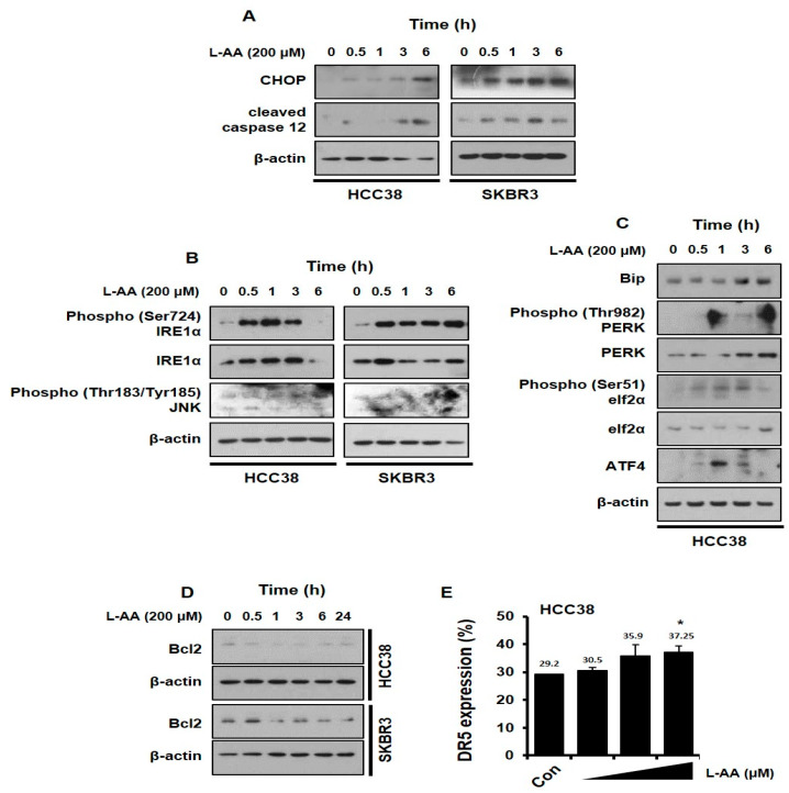
Figure 4.
L-AA increases endoplasmic reticulum (ER) stress in breast cancer cells. (A,D) HCC-38 or SKBR3 cells were treated with 200 μM of L-AA for indicated hours, and then western blotting was performed. β-actin was used as the loading control. (A) Expression levels of C/EBP homologous protein (CHOP) and cleaved caspase 12. (B) Protein levels involved in ER stress associated inositol-requiring endonuclease 1 (IRE1) signaling pathway. (C) Protein levels involved in ER stress-related PKR-like ER kinase (PERK) signaling pathway. (D) B-cell lymphoma 2 (Bcl2) expression levels (E) HCC-38 cells were treated with 200 μM of L-AA for 24 h and stained with anti-DR5 (death receptor 5) primary antibody (1:100) and –AlexaFluor-488 secondary antibody (1:1000). DR5-positive cells were detected by LSRFortessa flow cytometry. Data are shown as the mean of three independent experiments, and the error bars represent standard deviation (SD). * p < 0.05 versus the control group.

