Figure 6.
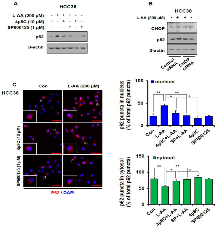
L-AA-induced IRE1–JNK–CHOP activation causes p62 accumulation in the nucleus. (A) HCC-38 cells were pre-treated with IRE1 signaling pathway inhibitors such as 4μ8C (10 μM) and SP600125 (1 μM) for 1 h and treated with L-AA (200 μM) for 6 h. Western blotting was performed with anti-p62, and β-actin was used as the internal control. (B) HCC-38 cells were transfected with CHOP siRNA, treated with L-AA (200 μM) for 6 h, and then western blotting was performed. (C) HCC-38 cells were pre-treated with the IRE1 signal pathway inhibitors, such as 4μ8C (10 μM) and SP600125 (1 μM), and treated with L-AA (200 μM) for 6 h. Cells were stained with anti-p62 (1 μg/mL) and –AlexaFluor-594 secondary antibody (1:200). Images were obtained using the FV10i confocal microscope, using the 40× objective; the scale bar indicates 50 μm. Following the experiment, three or more fields were obtained for each group, and p62 puncta were counted and averaged. This process was repeated three times, and the mean value of three experiments was statistically processed. The following graph shows the ratio of p62 puncta expressed in the cytosol and nucleus, based on 100% of total p62 puncta. Data are shown as the mean of three independent experiments, and the error bars represent standard deviation (SD). * p < 0.05 and ** p < 0.01 were considered statistically significant.

