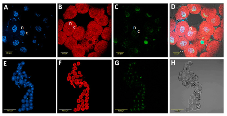
Figure 5.
Localization of Sodalis in B. trigonica bacteriocytes using FISH with Sodalis-specific probe. A, DAPI-stained bacteriocyte nuclei. B, Same photo as in A showing the localization of Carsonella, the primary symbiont of psyllids (red) in the bacteriocytes. C, Sodalis localization (green) inside bacteriocyte nuclei. D, Overlay of A, B and C under a bright field. E-G show a cluster of bacteriocytes with DAPI, Carsonella and Sodalis staining, respectively. H shows the same cluster of bacteriocytes under a bright field. n: nucleus; c: cell cytoplasm.

