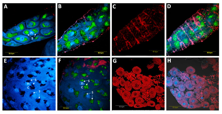
Figure 7.
Co-localization of Spiroplasma and CLso in the B. trigonica midgut using FISH. A, B, focal plans showing the localization of Spiroplasma (green) in BT midguts by FISH and DAPI-stained nuclei (blue). In these focal planes, some localization of CLso appears (red). C, the same portion of the midgut in B showing the localization of CLso (red). D, overlay of Spiroplasma, midgut nuclei and CLso under bright field. E-F, other views of Spiroplasma localization (green) and DAPI-stained nuclei (blue). E, Spiroplasma patches around the nuclei without any staining. F, Spiroplasma patches with staining. G, FISH using Carsonella (red) and Spiroplasma probes in bacteriocytes. Spiroplasma was not detected inside bacteriocyte cells. H is the overlay of Carsonella, Spiroplasma and DAPI-stained nuclei in bacteriocytes. n: nucleus; c: cellular cytoplasm. Arrows indicate, s: Spiroplasma.

