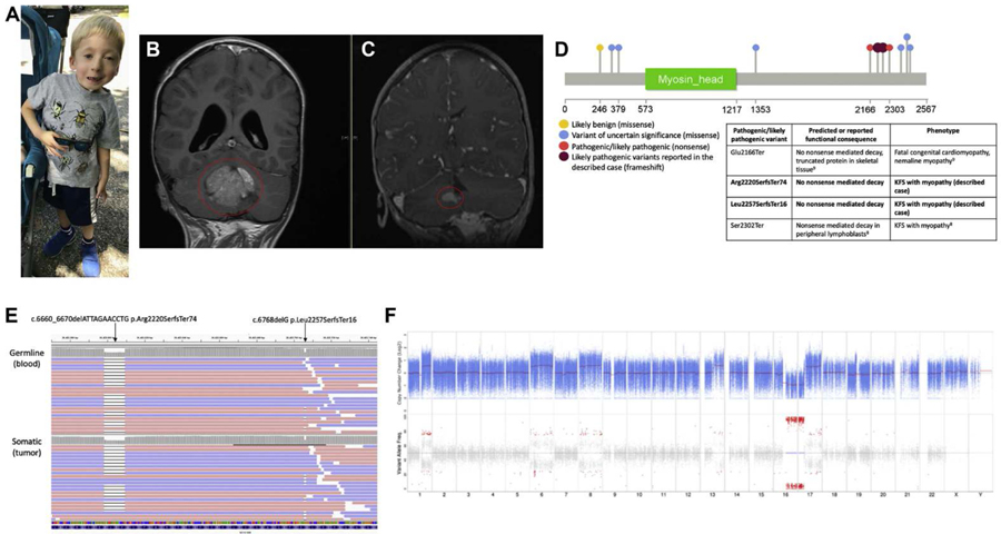
Fig. 1. Clinical presentation and molecular genomic findings.
(A) Full body image of the patient demonstrating clinical features of KFS and facial dysmorphism. (B) Magnetic resonance imaging of the brain with contrast, coronal T1 FLAIR at diagnosis demonstrating large homogenous cerebellar mass (circled). (C) Magnetic resonance imaging of the brain with contrast, coronal fast spoiled gradient-echo at recurrence demonstrating new nodules along the surface of the right cerebellum (circled). (D) Schematic of MYO18B variants reported in ClinVar with colors corresponding to variant interpretation: likely benign (yellow), blue (variant of uncertain significance), red tpathogenic/likely pathogenic). Likely pathogenic variants reported in the described patient are shown in dark red. A table describes the reported or predicted functional consequence of pathogenic/likely pathogenic variants and the associated patient phenotype. (E) Visualization of the MYO18B compound heterozygous variants suggestive of an in trans inheritance patterns. Top: Constitutional MYO18B reads from the peripheral blood aligned to GRCh37. Bottom: Somatic MYO18B reads from the tumor aligned to GRCh37. Reads are colored by read strand, red for positive strand and blue for negative strand. (F) Somatic copy member variation (CNV) analysis. Top: Tumor copy number relative to matched normal in 1og2 scale. Blue points represent log2 values based on sequence depth in 100-bp windows. Red lines indicate segmented CNV calls. Bottom: Tumor variant allele frequency for variants that are heterozygous in the normal. Points in red indicate significant loss of heterozygosity (LOH). The x-axis denotes the chromosome number.

