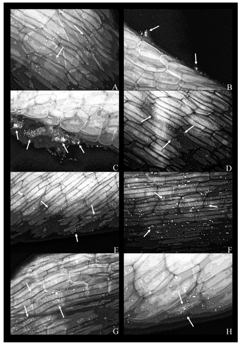
Figure 3.
Fluorescent micrographs of duckweed roots in phenol-supplemented (500 mg L−1) MS medium with bacterial strains: (A) 11—Lelliottia sp., bacterial cells scattered on the root surface(arrows); (B) 14—Klebsiella oxytoca, dense groups of bacterial cells near the root surface (arrows); (C) 14—K. oxytoca (63×), dense aggregates of bacterial cells (indicated by arrows) near the regions of the root destroyed by pressure (squashing); (D) 27—Serratia marcescens, bacterial cells organized along the lines between epidermal cells of the root (arrows); (E) 37—Hafnia alvei, bacterial cells organized on the root surface and along the lines of cell–cell boundaries (arrow); (F) 43—H. paralvei, bacterial cells on the root surface; (G) 51—S. nematodiphila, scarce bacterial cells on the root surface; (H) 51—S. nematodiphila, magnified 63× to show a typical microcolony on the root surface. Magnification: 40×, unless stated otherwise. Arrows: bacterial cells on the root surface.

