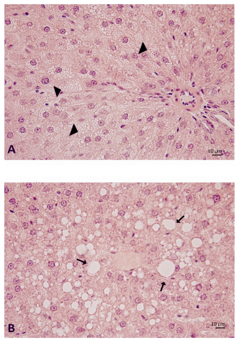
Figure 6.
Liver morphology of DIO rats. Sections of liver processed for haematoxylin and eosin staining to highlight the (A) microvesicular steatosis, mainly found in the periportal areas (arrow head), and (B) macrovesicular steatosis (arrow), in a centrilobular area where several scattered balloon cells can be often observed. Magnification 40×. Calibration bar: 10 μm. DIO—diet-induced obese rats.

