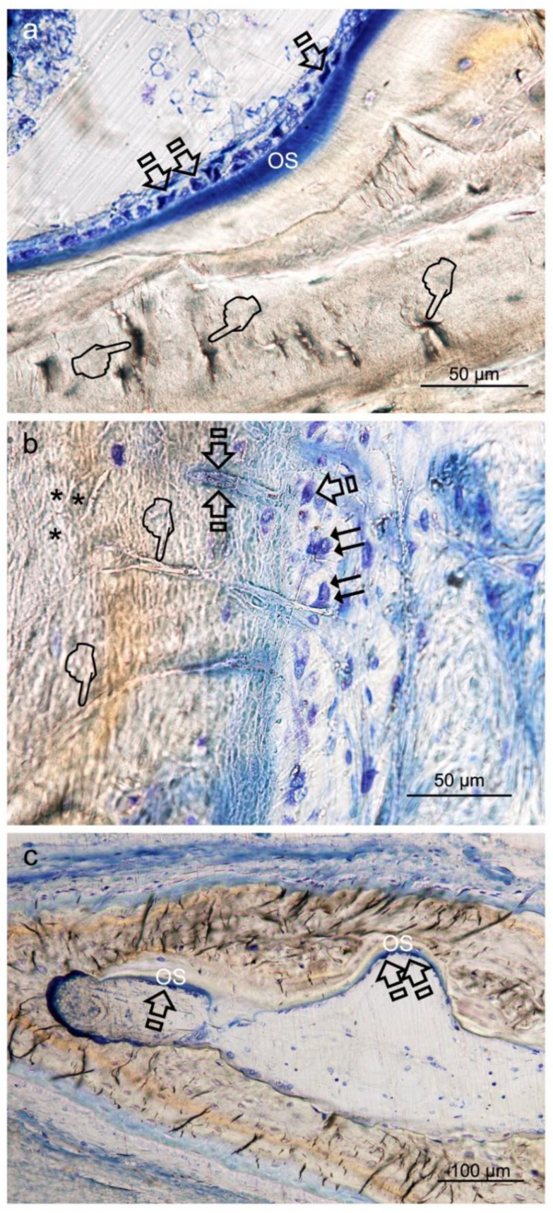
Figure 4.
Bone histology images obtained after using Zn-HOOC-Si (a), Dox-HOOC-Si (b) loaded membranes, and control (no membrane) (c), by coloration with toluidine blue to visualize mineralized bone, at 6 weeks of healing time. Single arrows point the presence of aligned osteoblasts, with typical cuboid shape (Figure 4a). Polarized osteoblast (Figure 4a,b) show part of the cell membrane in direct contact with the bone surface, unveiling many cytoplasmic processes, that achieve the newly deposited osteoid (OS). Double arrows are indicative of osteoclasts; pointers mean canaliculi and faced arrows, an osteocyte. Asterisks are located at zones of gap junctions (Figure 4b).

