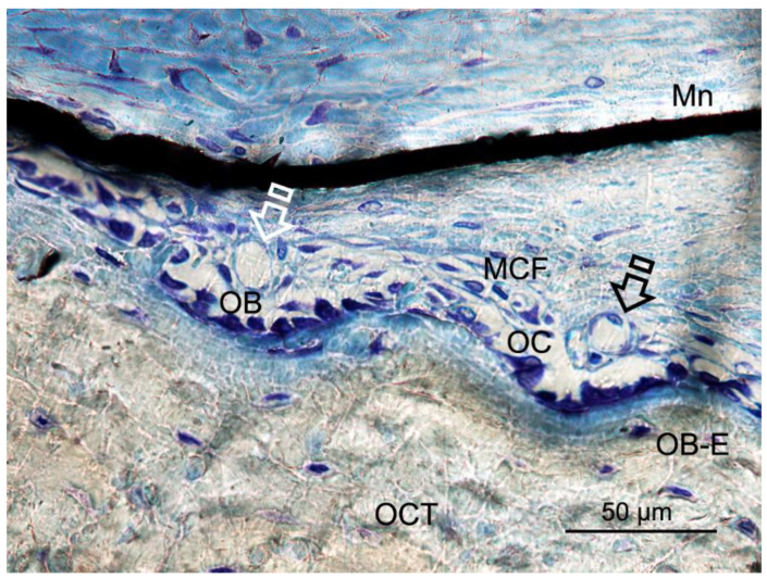
Figure 6.
Bone histology images obtained after using Zn-HOOC-Si loaded membrane (Mn), by dye with toluidine blue to visualize mineralized bone, at six weeks of healing time. Single arrows point the presence of blood vessels; both osteoblasts and osteoclasts are in close contact with marrow elements and the contiguous vasculature. Osteocytes (OCT), osteoblasts (OB), entrapped osteoblasts (OB-E), osteoclasts (OC) and macrophages (MCF) may be observed.

