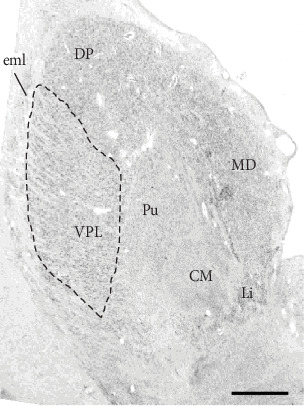
Figure 2.

Postmortem Tissue Sample stained for visualization and delineation of thalamus subnuclei. Our delineation of the VPL follows that of the standardized nomenclature of Feremutsch and Simma in Dewulf [105]. We sampled from the portion of VPL in coronal sections through the caudal half of the centromedian nucleus (CM). At these levels, VPL is rather conspicuous due to its relatively larger and more coarsely staining neurons compared to surrounding nuclei. Moreover, these neurons, especially in the more lateral extent of the nucleus exhibit a prominent diagonal arrangement. The lateral boundary of the VPL is the external medullary lamina of the thalamus; its superior border, the dorsal posterior (DP) nucleus with more homogeneously arranged cells; it medial boundary is the anterior portion of the pulvinar with conspicuously smaller, paler, and more homogeneously arranged cells. Abbreviations: CM: centromedian nucleus; DP: dorsal posterior nucleus; eml: external medullary lamina; Li: nucleus limitans; MD: mediodorsal nucleus; Pu: putamen.
