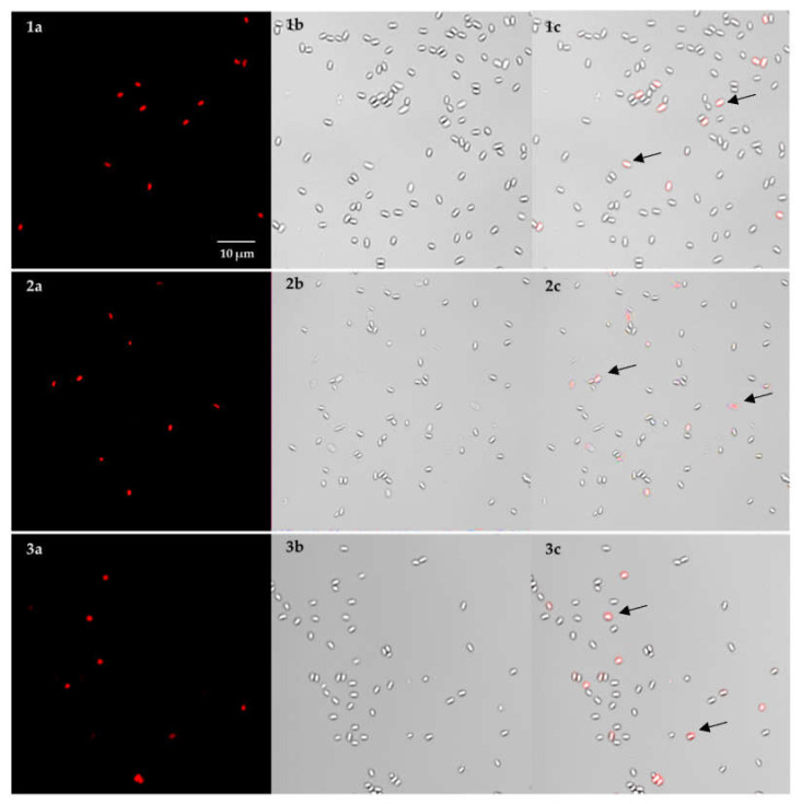
Figure 2.
Confocal laser scanning microscopy images of B. anthracis spores pre-treated with PMA before and after DNA extraction by two different methods. Micrographs were taken before (1) and after DNA extraction with the MasterPure Complete DNA and RNA Purification kit (2) and the QIAamp DNA Mini kit as comparison (3), respectively; (a) red channel (PMA dyed) to detect spores with compromised spore coat, cortex and membranes; (b) Bright field image, (c) Overlay of bright field and PMA signal; Scale bar: 10 μm. Arrows indicate spores with compromised spore coat, cortex, and membranes.

