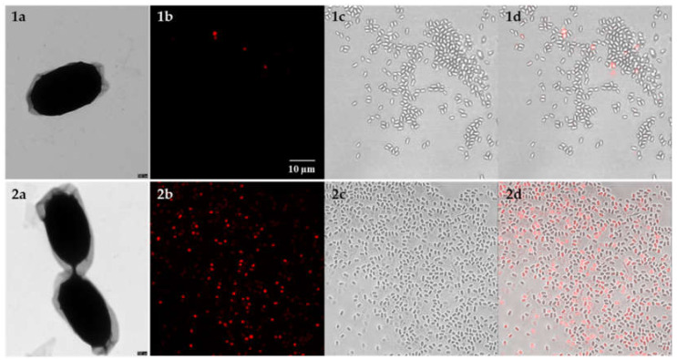
Figure 3.
Disruption of B. anthracis spores after bead-beating observed by TEM and fluorescence microscopy. Micrographs show untreated spores (1) and spores after bead-beating (2); (a) TEM of negatively stained B. anthracis Sterne spores (Scale bar: 100 nm); confocal laser scanning microscopy images of B. anthracis spores pre-treated with PMA before (1b–d) and after (2b–d) mechanical disruption of spores by bead-beating; (b) red channel (PMA dyed) to detect spores with compromised spore coat, cortex and membranes; (c) bright field image, (d) Overlay of bright field and PMA signal (scale bar for all light micrographs: 10 μm).

