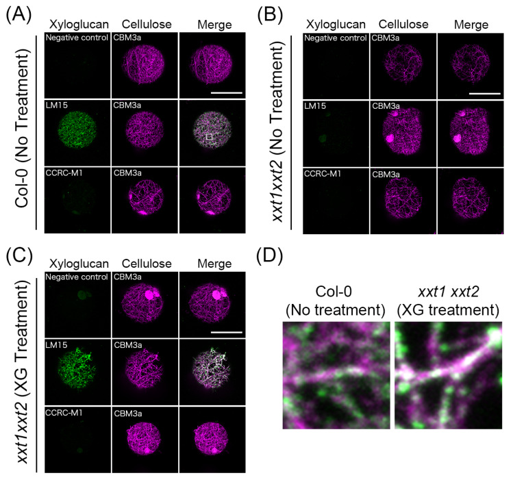
Figure 3.
Immunofluorescent labeling of xyloglucan and crystalline cellulose on the surface of Col-0 and xxt1 xxt2 mutant protoplasts. (A–C) Immunocytochemistry of protoplasts incubated in the regeneration medium lacking xyloglucan (A,B) or containing xyloglucan (C) for 24 h. Crystalline cellulose was stained with CBM3a, and xyloglucan was stained with LM15 or CCRC-M1. Distilled water was added instead of LM15 or CCRC-M1 as the negative control. (D) Magnified images of the white squares in (A) (left) and (C) (right). These images show the overlap between LM15 and CBM3a signals. Scale bar = 20 μm.

