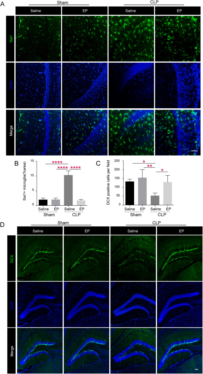
Fig. 2.

Ethyl pyruvate attenuates microglia activation and restores formation of neonatal neurons in septic mice. a Microglia activation of shamed or septic mice treated with EP (40 mg/kg) or vehicle (PBS) was assessed by Iba-1 immunofluorescence on postoperative day 12. Photomicrographs of CA1 areas of the hippocampus are shown infection activated microglia as noted by morphological changes on day12, including enlargement of cell bodies, unsmooth synapse, and so on, which was attenuated by EP treatment. Scale bar = 50 μm. b & c showing area percentage of activated microglia in the CA1 region and DCX positive cells in the dentate genus (DG) of septic mice hippocampus in Sham+Saline, Sham+EP, CLP + Saline and CLP + EP groups. Data presented as mean ± SEM (n ≥ 3 mice/ group) and compared by one-way ANOVA and post-hoc test, * P < 0.05, **P < 0.01, ****P < 0.0001. d Photomicrographs of representative DCX immunofluorescence (Doublecortin, green, neurogenesis) of shamed or septic mice treated with EP (40 mg/kg) or vehicle (PBS) in the DG areas on postoperative day 12. Scale bar =100 μm
