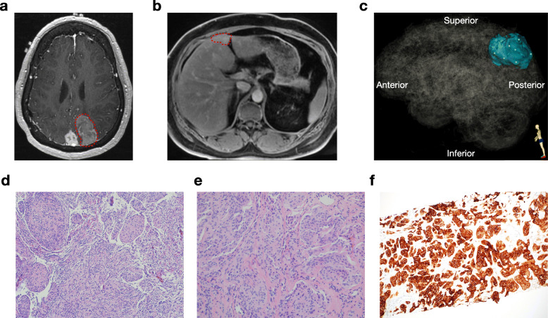
Fig. 1.
Pre-operative magnetic resonance imaging and histopathology of intracranial and metastatic meningioma. a Post-contrast T1 axial MR images show the enlarging left parieto-occipital meningioma (red circle), and a stable parasagittal meningioma. b Post-contrast liver MRI shows a metastatic lesion in segment IVb (red circle). c Three-dimensional stereotactic meningioma sampling map for 6 intracranial meningioma samples reconstructed from preoperative magnetic resonance imaging. d H&E stain of the intracranial meningioma sample (10x). e H&E stain of the liver metastasis core biopsy (20x). f SSTR2A immunohistochemistry of the liver metastasis core biopsy (10x)

