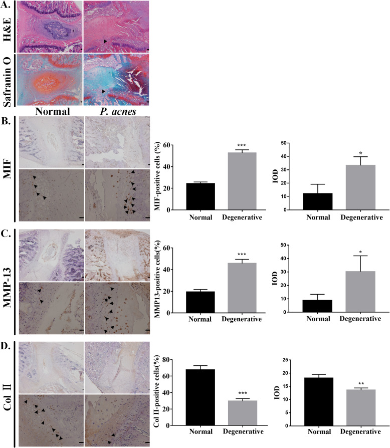
Fig. 2.
Changes in P. acnes-inoculated rat CEPs. a H&E staining and Safranin O fast green staining of the segment of P. acnes-inoculated intervertebral discs demonstrated the disappearance of the nucleus pulposus, endplate fracture (black arrow), and a disorganized annulus fibrosus. Immunohistochemistry of b MIF, c MMP-13, and d Col II showed that P. acnes induced CEP degeneration. The left side is the control group and the right side is the experimental group. The upper panels were amplified at × 40, while the lower panels were amplified at × 200. The black arrow represents immunopositive cells. Scale bar: 20 μm. *p < 0.05, **p < 0.01, ***p < 0.001

