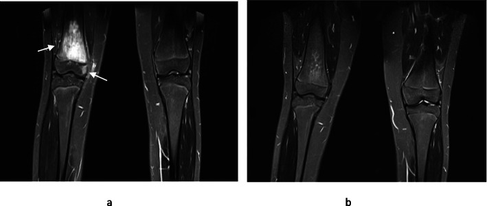
Fig. 2.
Pre and post pamidronate treatment MR images. Pre and post- pamidronate WB-MRI images of a 15 year old girl who presented with significant right knee pain and was diagnosed with CNO following a bone biopsy. Her symptoms resolved completely following four cycles of pamidronate. 2a – The coronal STIR MR image shows extensive high signal predominantly of the distal right femoral metaphysis consistent with intra-osseus oedema. A smaller area of the medial epiphysis is affected without features of cortical destruction or significant soft tissue component. 2b – Almost complete resolution of the metaphyseal high signal is in keeping with treatment response. The epiphyseal component is also no longer visible. In our exercise, the machine algorithm and panel of radiologists concurred that lesions resolved post treatment

