Figure 2.
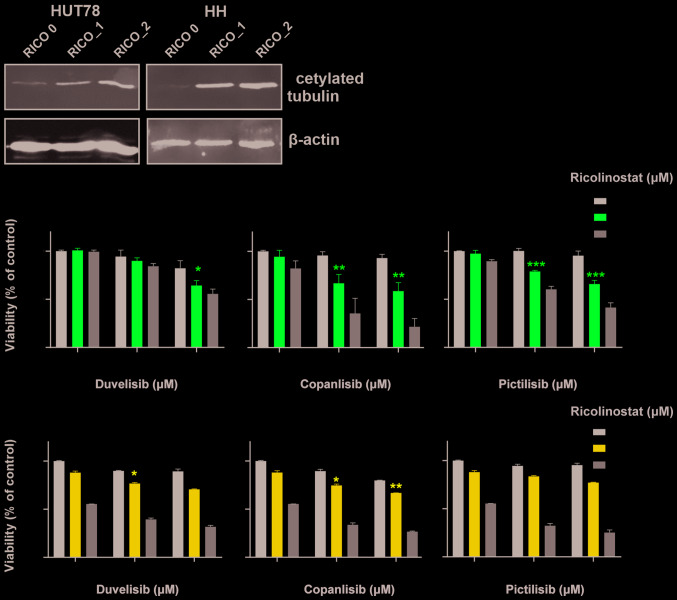
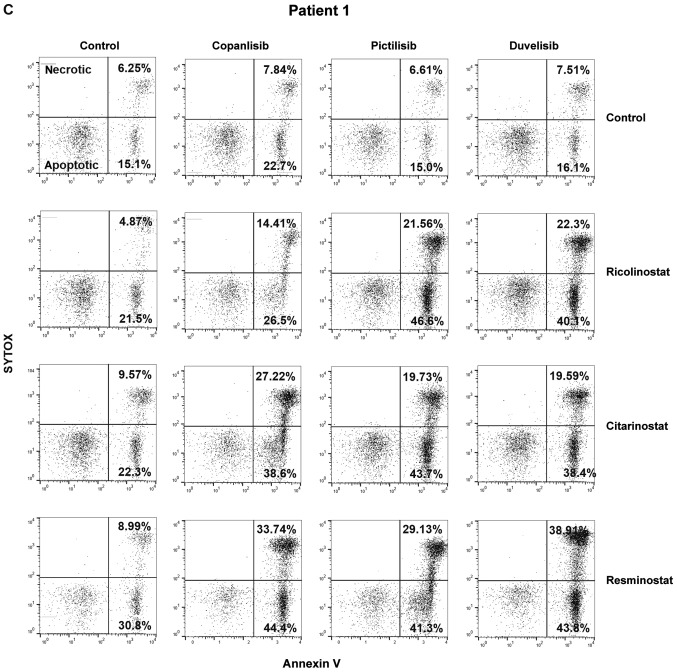
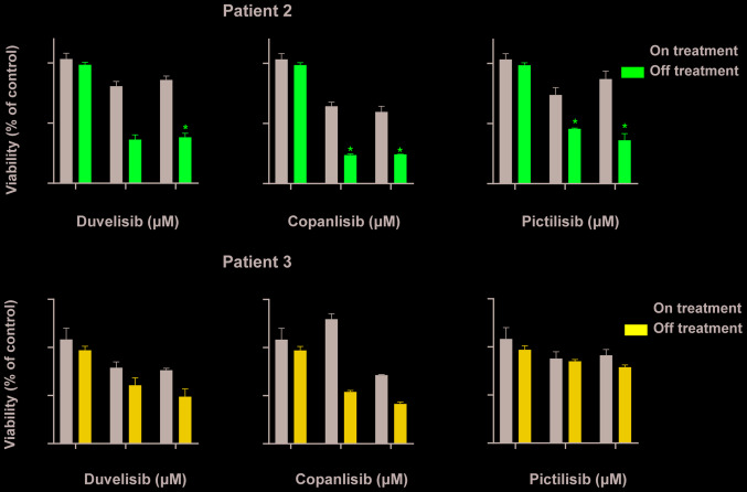
HDAC6 inhibitors sensitize CTCL cells to PI3K inhibitors. (A and B) Established CTCL cell lines (HUT78 and HH) were co-incubated for 48 h with increasing concentrations of the HDAC6 specific inhibitor, ricolinostat, and the following PI3K inhibitors: The non-selective inhibitor, copanlisib, the γ/δ-selective inhibitor, duvelisib, and the pan-inhibitor pictilisib. (A) Levels of acetylated tubulin (a hallmark of HDAC6 inhibition) were assessed by western blot analysis in whole-cell lysates of HUT78 and HH cell lines incubated with 0, 1 and 2 µM ricolinostat (RICO_0,1,2). β-actin was used as a loading control. (B) Cell death was assessed by flow cytometry using propidium iodide or SYTOX combined with Annexin V and proliferation was assessed by ATP quantification with Cell Titer Glo®. Statistical significance was assessed using the Mann-Whitney U test; *P<0.05, **P<0.01, ***P<0.001, ****P<0.0001. HDAC6 inhibitors sensitize CTCL cells to PI3K inhibitors. (C and D) HDAC6 inhibitors sensitize CTCL primary cells to PI3K inhibitors. PBMCs from a leukemic patient with CTCL were isolated using standard Ficoll centrifugation. (C) Cells were then co-incubated for 48 h in the presence of IL-2 and IL-15 stimulation with the HDAC 1/3/6-specific inhibitor, resminostat, or the HDAC6-specific molecules, ricolinostat and citarinostat (all at a 4 µM concentration), in combination with the PI3K inhibitors, copanlisib, pictilisib and duvelisib (1 µM). Cell viability was assessed by flow cytometry following Annexin V/SYTOX staining of CD3+ CD4+ cells. The figure shows the percentages of necrotic (upper panel) and apoptotic (lower panel) cells. (D) In addition, ATP activation was assessed using Cell Titer Glo®. HDAC6 inhibitors sensitize CTCL cells to PI3K inhibitors. (E and F) HDACi-based therapy sensitizes primary CTCL cells to PI3K inhibitors. PBMCs from leukemic patients with CTCL ‘on and off’ therapy with (E) the HDAC pan-inhibitor, vorinostat, and (F) the HDAC1/3/6 specific inhibitor, ricolinostat, were then co-incubated for 48 h in the presence of IL-2 and IL-15 with 1 and 2 µM PI3K inhibitors, copanlisib, pictilisib and duvelisib. Cell viability was assessed by ATP quantification using Cell Titer Glo®. Statistical significance was assessed by two-way ANOVA with multiple comparisons; *P<0.05. RICO, ricolinostat; CITA, citarinostat; RESMI, resminostat; CTCL, cutaneous T-cell lymphoma; HDAC6, histone deacetylase 6.


