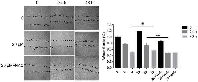
Figure 4.
MDA-MB-468 cells were seeded in 6-well plates and allowed to reach a confluent monolayer. The cells were incubated with various concentrations of propofol for 0, 24 or 48 h. Cells were also incubated with 20 µM propofol + NAC for 48 h. (A) Wound healing assay and (B) analysis performed relative to the starting wound width at 0 h. Data are represented as the mean ± SD (n=3). #P<0.05 (0 vs. 20 µM propofol for 48 h) **P<0.01 (20 µM propofol vs. 20 µM propofol + NAC for 48 h). NAC, N-acetyl-L-cysteine.

