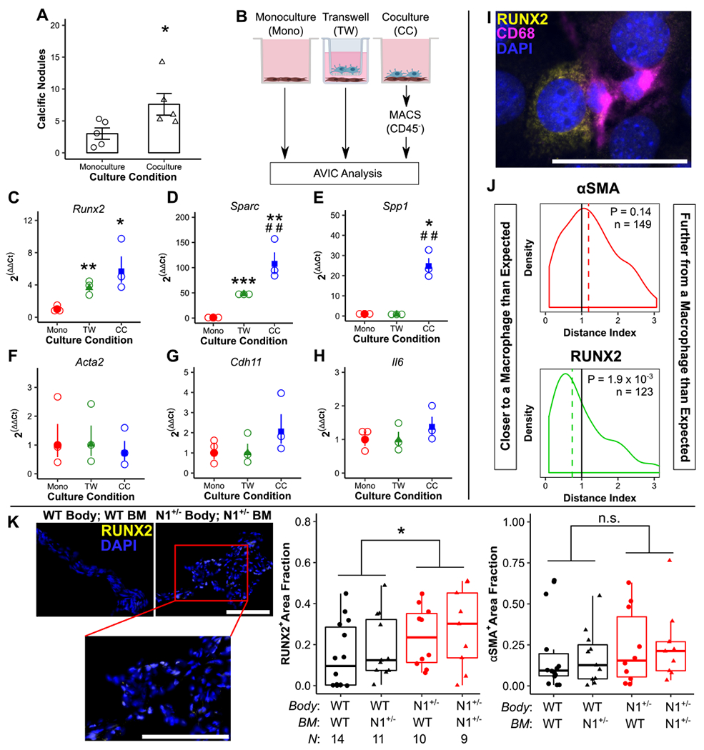
Figure 4. Macrophages promote osteogenic but not dystrophic calcification of aortic valve interstitial cells.

Cocultures of aortic valve interstitial cells (AVICs) with bone marrow-derived macrophages (BMMs) were assayed for calcific nodule formation (A, N = 5). AVICs cultured in monoculture (Mono), Transwell culture with BMMs (TW), or direct coculture with BMMs (CC) (B) were assayed for transcription of markers of osteogenic (C-E) and dystrophic (F-H) calcification (N = 3). Images of cocultures stained for RUNX2, CD68, and DAPI (I) were analyzed by a Monte Carlo-assisted simulation to calculate expected distance and distance index between activated AVICs and BMMs (J). Bone marrow transplanted wild-type (WT) and Notch1+/− (N1+/−) mice were stained for RUNX2 and αSMA expression (K). Scale bars = 200 μm (A, K) and 1 mm (I). All summary data represent mean ± s.e.m. Data were analyzed by Mann Whitney U test (A), one-way ANOVA followed by two-tailed t tests with Holm-Sidak corrections on untransformed ΔCt values (C-H), one sample Wilcoxon Signed-Rank test on log-transformed data (L), or two-way aligned rank transformed ANOVA (K). *P < 0.05, **P < 0.01, ***P < 0.001 from monoculture AVICs (A-H) or between genotypes (K); ##P < 0.01 from Transwell AVICs. N = biological replicates (B-K) or activated AVICs across 4 biological replicates (L).
