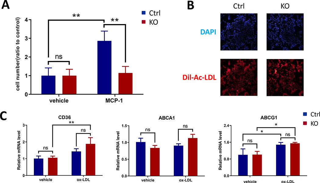
Figure 4. Bmal1 deficient macrophages showed reduced migration potential and unaltered lipid handling in vitro.
(A) Cells were placed in Transwell upper chambers and chemotaxis was assessed with or without exposure to 10ng/ml MCP-1, the KO cells showed significantly reduced migration to MCP-1. (B) Cells were treated with 10μg/ml DiI-Ac-LDL for 4h and uptake was assessed. Representative fluorescence images showed no difference between Ctrl and KO cells. DAPI staining was used as a control. (C) Cells were treated with or without 50μg/ml ox-LDL for 24h, real-time RT-PCR showed no difference between Ctrl and KO cells for CD36, ABCA1 and ABCG1 expression. (2-way ANOVA with Tukey’s multiple comparisons test, n=3, * p<0.05, ** p<0.01, ns, no statistical difference)

