Figure 2:

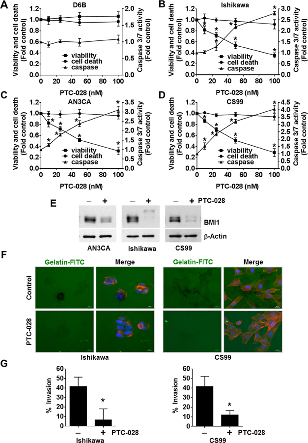
PTC-028 inhibits endometrial cancer cell viability and invasion. (A) D6B, (B) Ishikawa, (C) AN3CA and (D) CS99 cells were treated with increasing concentrations of PTC-028 for 48h and cell viability, cytotoxic cell death and caspase 3/7 activity was evaluated using the ApoTox-Glo Triplex assay. Data are mean ± S.D. of three independent experiments performed in triplicate. *P<0.05 when comparing with respective vehicle treated control by two-way ANOVA. (E) AN3CA, Ishikawa and CS99 cells were treated with 50 nM PTC-028 for 48h. Expression of BMI1 was determined by immunoblotting. (F) Representative images of Ishikawa and CS99 cells plated on Oregon Green® 488 Gelatin coated coverslips and treated with 20 nM PTC-028 for 24h (CS99) or 36h (Ishikawa). The cells were fixed and stained with Alexa Fluor® 555 Phalloidin and mounted in Vectashield mounting medium containing DAPI. (G) Quantification of invaded cells were counted from ~100 cells per treatment group as represented in Fig. 2F. Data are mean ± S.D. of three independent experiments performed in triplicate. *P<0.05 when comparing with respective vehicle treated control by Student’s t-test.
