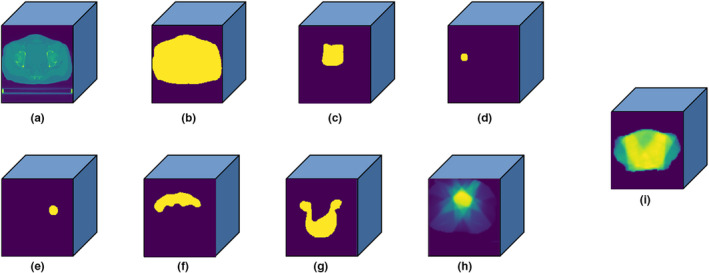
Fig. 1.

Three‐dimensional (3D) matrix (128 × 128 × 128) of (a) computed tomography images; (b) body; (c) bladder; (d) right femoral head; (e) left femoral head; (f) small intestine; (g) planned target volume; (h) beam configurations, and (i) 3D dose distribution.
