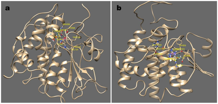
Figure 5.
Major sub-groups of GST and its binding mode with substrate GSH are depicted in this picture (a); (b) show the stereo-view of GSH binding with cyGST X6 and X7, a representative structure for S and C GST. S type GST resides in GSH between helix 1,4, and 8 with the involvement of beta strand 2. Similarly with C type GST, the GSH interaction was observed with helix 1, and 7 and beta strand 1.

