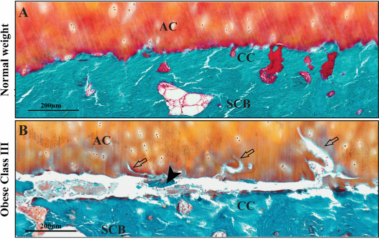
Figure 1.
Representative images of horizontal fissures in the osteochondral units from OA patients with normal weight and morbid obesity. In patients with normal weight (A), cartilage is firmly attached on calcified cartilage (CC). In patients with obese class III (B), a horizontal fissure is observed at the osteochondral interface between the articular cartilage (AC) and subchondral bone (SCB). Free bone debris (black arrowhead) and cartilage erosion (empty arrow) are presented within the fissure.

