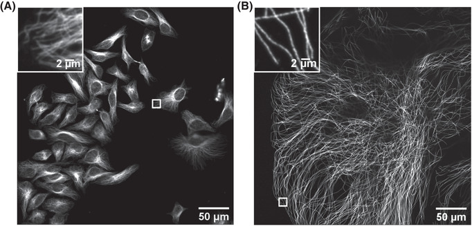
Figure 4.

Imaging of HeLa cells with immunostained microtubules before and after expansion. Imaging was performed on an Andor spinning‐disk confocal microscope with a 40×, numerical aperture (NA) 1.15 water‐immersion objective. (A) Confocal image of HeLa cells with immunostained microtubules, imaged at a single xy plane at the bottom of the cells. The inset in the upper left zooms in on the small box at the middle right. (B) Confocal image of a ∼4.5× linearly expanded HeLa cell with immunostained microtubules, imaged at a single xy plane at the bottom of the cell. The inset in the upper left zooms in on the small box at the bottom left. Scale bars in (B) indicate post‐expansion scales.
