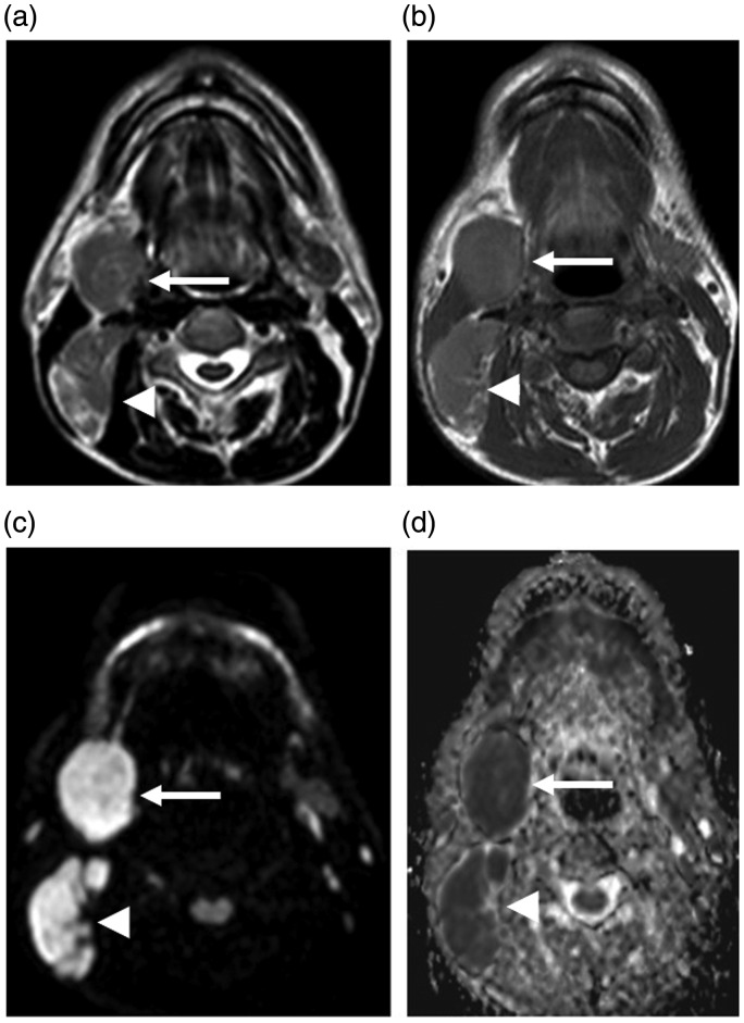
Figure 1.
Fifty-six year-old male with a history of right cervical mass. A well-defined right submandibular mass lesion with intermediate signal intensity on T1WI (b) and T2WI (a) (arrows). Enlarged right cervical lymph nodes are also observed (arrowheads). They show a bright signal on diffusion-weighted MRI (c) and have an ADC value (d) of 0.5 × 10⁻³ mm2/s. Pathological assessment shows classical Hodgkin lymphoma with mixed cellularity.

