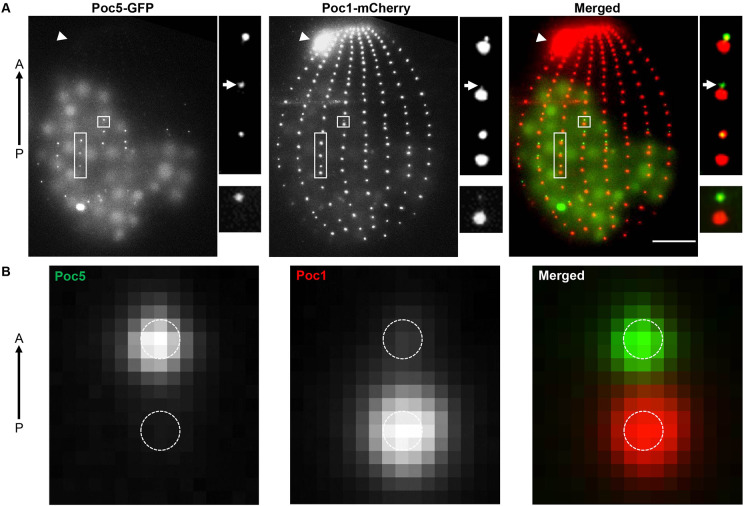
Fig. 2.
Endogenously tagged Poc5–GFP localizes to assembling BBs. (A) Live-cell imaging of C-terminally tagged Poc5–GFP relative to Poc1–mCherry (BB marker) with the anterior (A)–posterior (P) axis indicated with an arrow. Poc5–GFP localizes to a subset of cortical row BBs (highlighted with boxes) but is not detected in the mature oral apparatus (labeled with arrowheads). Scale bar: 10 µm. Upper right panels are representative regions of cortical rows containing three BB pairs. Poc5–GFP localizes to the anterior (assembling) BBs and is absent in the posterior (mature) BBs. Poc5 incorporation can precede that of Poc1 (labeled with arrows). Small (1.1 µm×1.2 µm) panels show the representative BB pair used for image averaging in B. (B) Image averaging of Poc5–GFP and Poc1–mCherry signals across 58 cortical row BB pairs reveals Poc5–GFP localizes exclusively at assembling BBs. The BB scaffolds are approximated with 200 nm diameter dashed circles based on the center of the Poc1–mCherry signal, reflecting the average diameter of a Tetrahymena BB.

