Fig. 3.
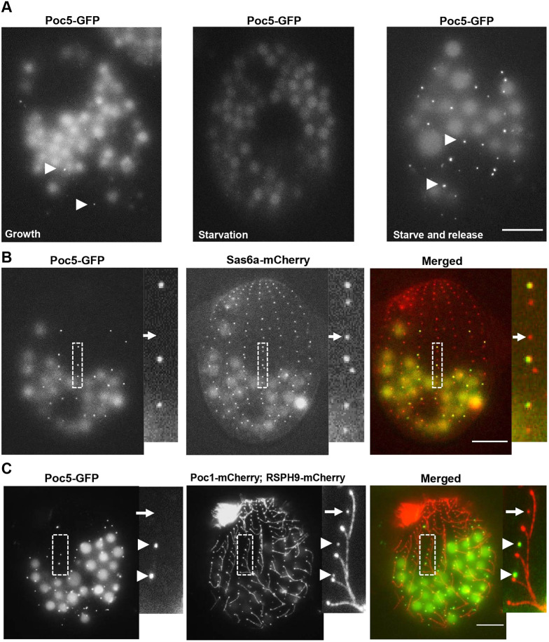
TtPoc5 is enriched during BB assembly and removed prior to cilia formation. (A) Live-cell imaging of endogenously tagged, C-terminal Poc5–GFP during logarithmic growth, in starvation and after release back into growth medium (starve and release). During logarithmic growth, BB assembly occurs throughout the cell cycle and low levels of Poc5–GFP signal are detected at a single moment (indicated with arrowheads). In starvation, cells are arrested in G1 preventing new BB assembly and Poc5–GFP is not detectable. Overnight starvation followed by release into growth medium (stimulating new BB assembly) enriches the Poc5–GFP signal (marked with arrowheads). (B,C) Live-cell imaging of starved and released cells with representative sections of cortical rows (marked with dashed boxes) expanded in the smaller panels. (B) Endogenous Poc5–GFP was co-expressed with Sas6a–mCherry (early marker of BB cartwheel structure) to assess the timing of TtPoc5 BB incorporation. Sas6a–mCherry labels all BBs whereas Poc5–GFP is only at the anterior BB in a pair. The Poc5–GFP signal always coincides with the Sas6a–mCherry signal, therefore TtPoc5 BB incorporation does not precede that of Sas6a. (C) Endogenous Poc5–GFP was co-expressed with Poc1–mCherry (BB marker) and RSPH9–mCherry (evenly labels ciliary axonemes) to assess the timing of Poc5 removal from maturing BBs. Poc5–GFP removal precedes cilia formation since Poc5–GFP-positive BBs are not ciliated (indicated with arrowheads) and maturing, non-ciliated BBs with detectable Poc1–mCherry (highlighted with an arrow) can be devoid of Poc5-GFP signal. Scale bars: 10 µm.

