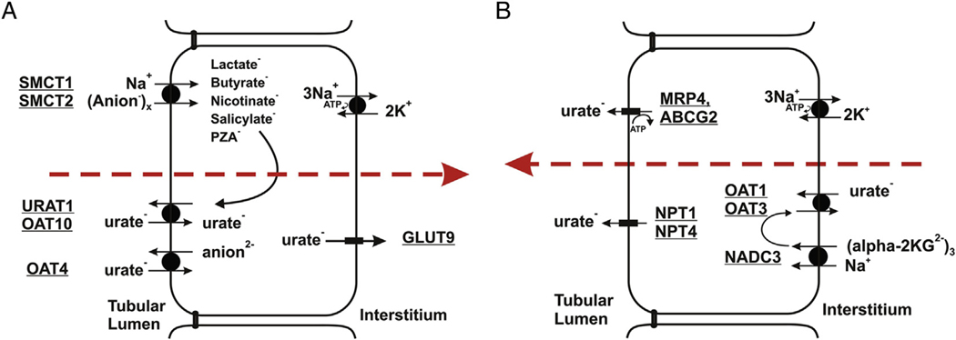
Figure 1.
Transport pathways for urate in proximal tubule cells. (A) Urate reabsorption. Sodium-dependent anion transport by SMCT1 and SMCT2 increases intracellular concentrations of monovalent anions that exchange with luminal urate (URAT1/OAT10). OAT4 appears to exchange urate with divalent anions. GLUT9 is the exit pathway for urate at the basolateral membrane. (A) Urate secretion. Urate enters the cell at the basolateral membrane by exchange with α-ketoglutarate, mediated by OAT1 and OAT3. At the apical membrane, urate is secreted by MRP4, ABCG2, NPT1, and/or NPT4. Figure is copyright Annual Reviews and is reproduced from Mandal and Mount.7

