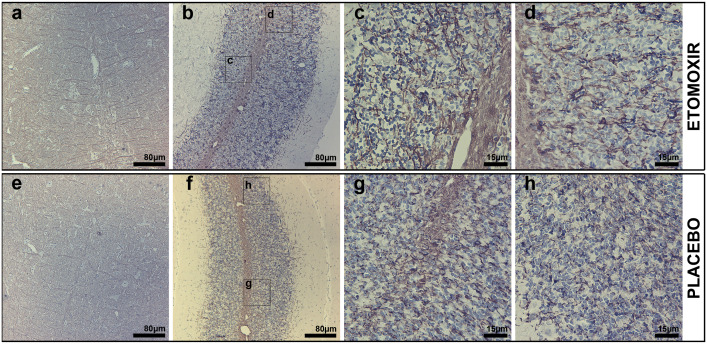
Fig 5. Immunoperoxidase microscopy of myelin basic protein (MBP) in coronal brainstem and cerebellum of etomoxir-treated rats (a-d) and placebo rats (e-h).
The etomoxir-treated group shown to possess higher MBP-labeling in the brainstem visualized as fibrous structures laterally protruding from nucleus raphe magnus and medial longitudinal fascicle compared to the group receiving placebo (a and e). Likewise, in the cerebellar white matter of etomoxir-treated rats, MBP-labeling was markedly more intense compared with placebo-receiving rats (b and f). MBP axons ensheathing in etomoxir-treated rats was labeled in the form of fibrous bundles in the cerebellar white matter, granular and molecular cerebellar layers, whereas in placebo animals MBP-labeled fibrous bundles were sparse and labeling pattern was more (c,d,g and h).

