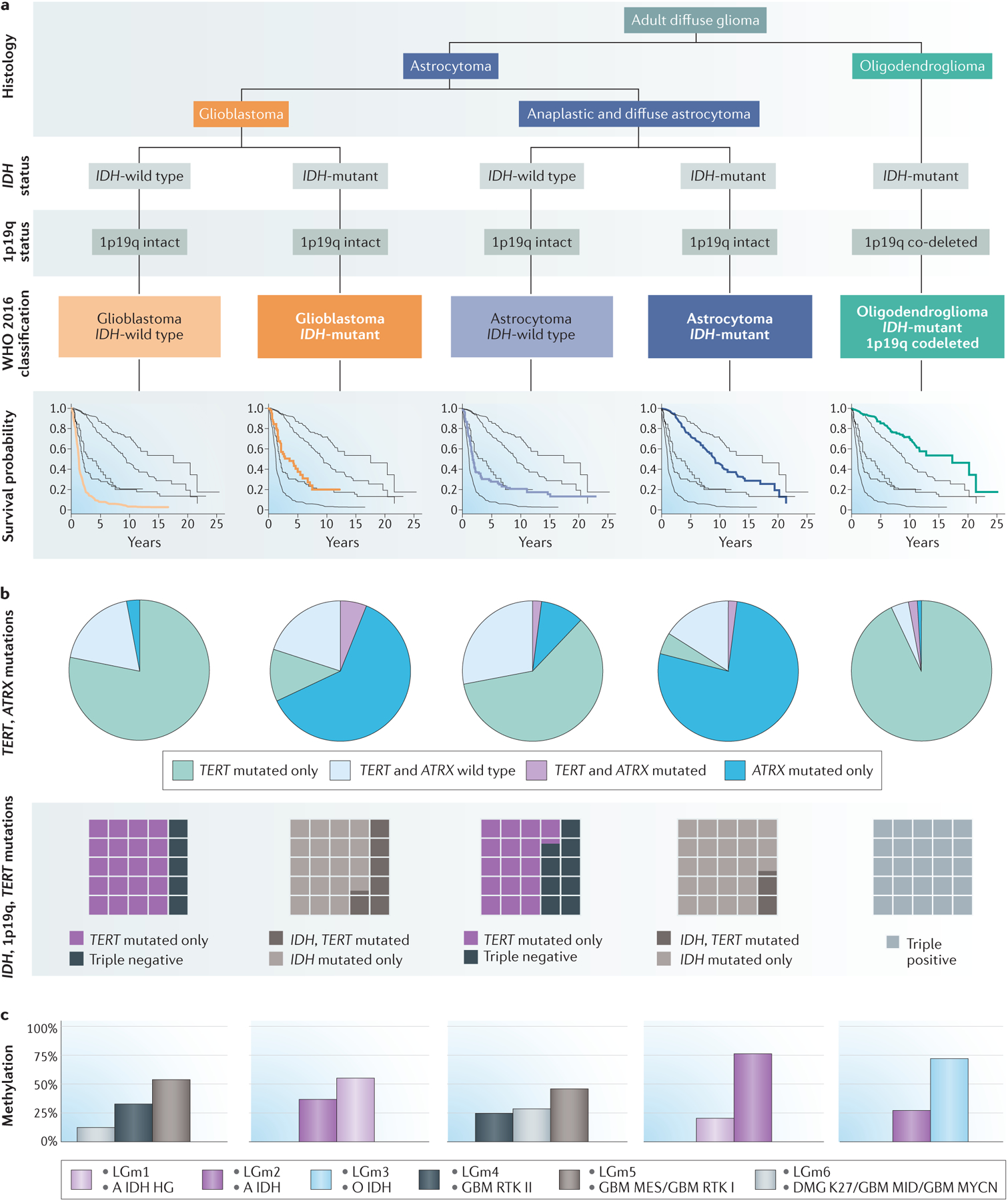
Figure 1. Overview of key molecular subtypes in the WHO 2016 classification of newly diagnosed adult diffuse glioma.

a | Histological assessment integrated with molecular diagnosis based on IDH mutation and 1p19q co-deletion status defines the five subtypes of adult diffuse glioma included in the WHO 2016 classification12,26. Kaplan–Meier survival curves for each type are based on data from 1989–201237. b | For each WHO 2016 disease subtype, telomerase-related mutation frequencies are depicted in pie charts showing the proportion of tumours with mutations in TERT and/or ATRX (green, TERT mutations only; blue, ATRX mutations only; purple, mutations in both TERT and ATRX; light blue, wild type TERT and wild type ATRX)37. Waffle plots show the relative proportions of IDH-mutant, TERT-mutant and 1p19q co-deleted tumours in each WHO 2016 disease subtype27. c | Graphs showing methylation classifications for each WHO 2016 subtype. The methylation categories depicted49 overlap to a great extent with those identified in a subsequent study46 (pink, LGm1 and A IDH HG; purple, LGm2 and A IDH; light blue, LGm3 and O IDH; black, LGm4 and GBM RTK II; gray, LGm5 and GBM MES/GBM RTK I; brown-grey, LGm6 and DMG K27/GBM MID/GBM MYCN). WHO 2016 classification from REFS.12,26. Survival data and telomerase mutation data from REF.37 with additional mutation data from REF.27. Methylation profile data from REFS.46,49.
