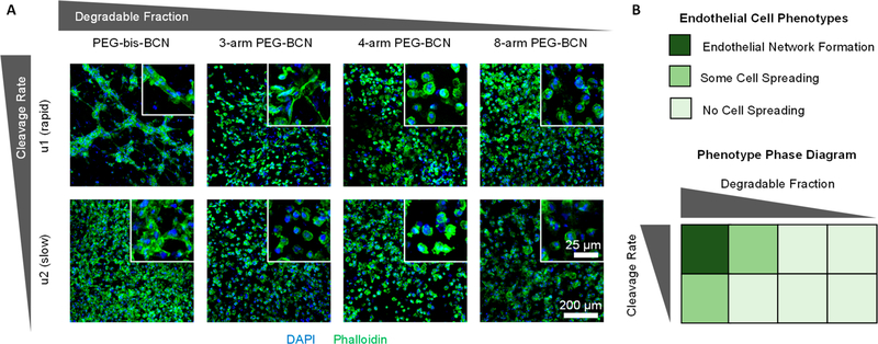
Figure 4.
(A) Confocal microscopy of brain microvascular cells embedded in ELP hydrogels reveals that endothelial network formation depends on both the chemical bond cleavage kinetics and the degraded hydrogel architecture. Insets are higher magnification images to assess cell spreading. Representative images were selected from 3 biological replicates. Blue: nuclei (DAPI), Green: F-actin (phalloidin). (B) “Phase diagram” summarizing how endothelial cell phenotype is affected by chemical bond cleavage rate and degraded hydrogel architecture.

