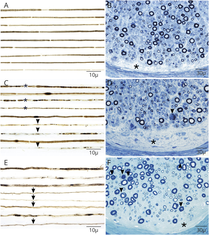
Figure 2. Nerve pathology of contactin-1 IgG neuropathy is distinct from classic-CIDP.
Three example patients with contactin-1 autoimmunity are shown (A–F). Closely approximated osmium-fixed teased nerve fibers from a fascicular biopsy demonstrate widened nodes of Ranvier with demyelination (A) compared with 2 sural nerve biopsies (C and F) where axonal injury is prominent including with early (asterisk) and late (arrowhead) degenerative stages including with empty nerve fibers (arrow) (E). Semithin sections stained with toluidine blue in all patients had distinct prominent subperineural edema (asterisk) without onion bulb formation as is typical in CIDP. Axonal degeneration was most prominently seen in (D, F-arrowheads). CIDP = chronic inflammatory demyelinating polyradiculoneuropathy.

