
Figure 1. CT of the head findings over time.
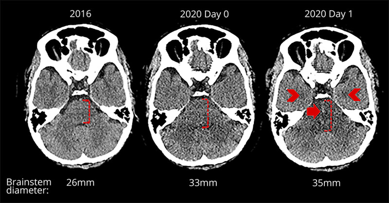
Axial CT head images on different dates. From left to right, premorbid previous CT performed in 2016, 2020 day 0 admission CT, and day 1 follow-up CT. Early admission CT demonstrates subtle new swelling of the brain stem, and the follow-up CT 1 day later shows progression of the swelling with new central hemorrhagic foci (closed arrow) and symmetrical hypodensities in both amygdalae (chevrons). On day 1 of the follow-up CT, there was also hypodensity in both thalami and dorsolateral putamina (not shown).