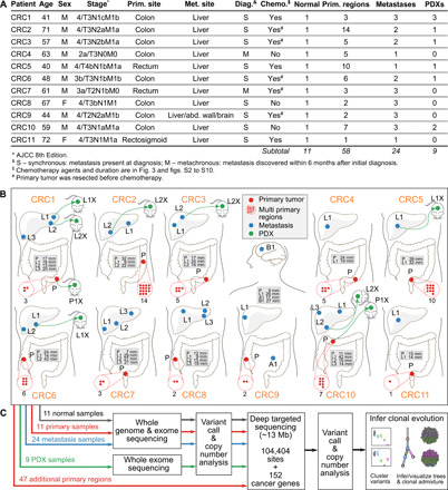
Fig. 1. Overview of patient cohort, samples, and study design.

(A) Summary of the clinical data. (B) Locations of the primary tumors and distant metastasis sites for all 11 patients. Sample names are prefixed by a letter representing the site of tumor (P, primary; L, liver metastasis; A, abdominal wall metastasis; and B, brain metastasis). Tumor diameters are in the gray boxes. The number of red dots inside red dashed circles indicates the total primary regions for the corresponding primary tumors. Several primary and metastasis samples were implanted into immunodeficient mice yielding PDX tumors (named with suffix letter X with a green dotted arrow linked to the parental patient sample). (C) Sequencing and clonal evolution analysis.
