Abstract
Primary sclerosing cholangitis (PSC) is characterized by biliary damage and fibrosis. Multidrug resistance-2 gene knockout (Mdr2−/−) mice and PSC patients have increased histamine (HA) levels (synthesized by l-histidine decarboxylase, HDC) and HA receptor (HR) expression. Cholestatic HDC−/− mice display ameliorated biliary damage and hepatic fibrosis. The current study evaluated the effects of knockout of HDC−/− in Mdr2−/− mice (DKO) on biliary damage and hepatic fibrosis. WT, Mdr2−/− mice and homozygous DKO mice were used. Selected DKO mice were treated with HA. We evaluated liver damage along with HDC expression and HA serum levels. Changes in ductular reaction were evaluated along with liver fibrosis, inflammation and bile acid signaling pathways. The expression of H1HR/PKC-α/TGF-β1 and H2HR/pERK/VEGF-C was determined. In vitro, cholangiocyte lines were treated with HA with/without H1/H2 inhibitors before measuring: H1/H2HR, TGF-β1 and VEGF-C expression. Knockout of HDC ameliorates hepatic damage, ductular reaction, fibrosis, inflammation, bile acid signaling and H1HR/PKC-α/TGF-β1 and H2HR/pERK/VEGF-C signaling. Reactivation of the HDC/HA axis increased these parameters. In vitro, stimulation with HA increased HR expression and PKC-α, TGF-β1 and VEGF-C expression, which was reduced with HR inhibitors. Our data demonstrate the key role for the HDC/HA axis in the management of PSC progression.
INTRODUCTION
Primary Sclerosing Cholangitis (PSC) is characterized by biliary damage, inflammation and hepatic fibrosis 1, 2. Typically, increased peribiliary inflammation leads to sclerosis of bile ducts eventually resulting in hepatic fibrosis and cirrhosis 3, 4. PSC can target specific subpopulations of the biliary tree including small and large bile ducts; however, large ductal PSC is more common (~70% of cases) and patients are at a greater risk for developing cholangiocarcinoma 5, 6. Coupled with impaired liver function, patients with PSC also suffer from severe side effects including chronic itching (pruritus) and fatigue 3, 4. Possible causes of pruritus are increased levels of circulating bile acids and elevated inflammatory cytokines 3. Treatment for PSC is currently lacking and the mechanisms of action which drive PSC are not fully understood.
Histamine is the major mediator released from activated mast cells (MCs) and interacts with one of four G-protein coupled receptors, H1-H4 histamine receptor (HRs) 7, 8. Histamine is produced following the conversion of histidine to histamine by the enzyme, l-histidine decarboxylase (HDC) which is expressed primarily by cholangiocytes in the liver 9, 10. Once released, histamine is either used or rapidly stored within MCs. Our previous study has demonstrated a differential regulation of H1 and H2HR in the proliferation of small and large cholangiocytes via Ca2+/PKC-α and cAMP/pERK signaling, respectively 10. Inhibition of HDC using either pharmacological compounds (α-methyl-DL-histidine dihydrochloride, α-Me) or genetic knockdown (global HDC−/− mice) results in reversal of biliary damage and hepatic fibrosis during cholestatic liver injury induced by extrahepatic bile duct ligation (BDL) 11. In addition, depletion of HDC reduces inflammation and angiogenesis in this chronic model of damage. HDC−/− mice subjected to a high fat, high sugar diet also showed amelioration of ductular reaction and liver fibrosis 12.
Hepatic fibrosis is a hallmark feature of PSC and a key regulator of this process is transforming growth factor (TGF)-β1 1, 2, 13. Several studies have demonstrated that TGF-β1 levels increase in Mdr2−/− mice 1, 2 and in human PSC 14. In addition, we have shown that inhibition of MC-derived histamine using cromolyn sodium decreases biliary damage and TGF-β1 expression and secretion, suggesting that histamine regulates TGF-β1 1. The cellular source of TGF-β1 is controversial; however, recent work has shown that cholangiocytes may provide a significant amount of TGF-β1 to the liver during PSC progression that mediates biliary senescence and liver fibrosis through autocrine and paracrine mechanisms, respectively 1, 2, 14. It is not fully known how histamine or histamine receptors may regulate TGF-β1 or what the relationship between these factors might be.
Vascular remodeling and alterations in the vascular bed are features of PSC and contribute to overall liver damage. There is evidence that links new blood vessel formation around bile ducts in response to chronic inflammatory liver diseases such as PSC 15. New vessel growth may be key in remodeling the tissue after injury, as well as delivering oxygen and other metabolic nutrients to damaged, hypoxic areas. In a study from 2014, we found that histamine regulates vascular endothelial growth factor (VEGF) signaling 11. In HDC−/− mice, VEGF expression and secretion were significantly reduced along with a downregulation of hypoxia inducible factor-α (HIF-1α), which is a secondary signaling molecule between histamine and VEGF 11. Further, in mice lacking MCs (decreased histamine synthesis) or in Mdr2−/− mice treated with cromolyn sodium, there is a reduction in vascular cell activation and VEGF signaling 1, 13. The goal of our current study is to demonstrate the critical contribution of the HDC/histamine axis to PSC progression via modulation of TGF-β1 and VEGF-C signaling via specific HR signaling.
MATERIALS & METHODS
Materials:
All reagents were obtained from Sigma-Aldrich, Co. (St. Louis, MO) unless otherwise indicated. Reagents and media for cell culture were obtained from Invitrogen Corporation (Carlsbad, CA) 2, 16. Total RNA was isolated from total liver tissues and purified cholangiocytes by the TRI Reagent from Sigma Life Science and reverse transcribed with the Reaction Ready First Strand cDNA Synthesis kit (Qiagen, Valencia, CA) 2, 16.
Animal Models:
Animal experiments were performed according to protocols approved by the Baylor Scott & White IACUC Committee and were performed in Temple, Texas prior to relocation to Indiana University School of Medicine. FVB/NJ, Balb/c and multidrug resistance 2 knockout (Mdr2−/−; strain FVB/NJ) mice were purchased from Jackson Laboratory (Sacramento, CA). HDC knockout mice (HDC−/−; strain Balb/c) were obtained from Dr. J. R. Goldenring (Vanderbilt University) and are currently breeding in our facility. The Mdr2−/− and HDC−/− mouse colonies have been established at our animal facility 1, 2, 12.
Generation of homozygote DKO mice:
Background crossed FVB/NJ x Balb/c (FVB/Balb) mice were first generated in order to evaluate strain differences between our animal groups and were subsequently utilized and referred to as wild-type (WT) control throughout the study. Mdr2−/− and HDC−/− mice were crossed until a homozygous Mdr2/HDC double knockout (DKO) strain was obtained. In the first cross only heterozygotes were obtained, as predicted. Selected heterozygotes from separate litters were then selected for breeding and a third generation was obtained that contained both male and female homozygotes (6.25% of mice bred were DKO homozygote). All genotyping was confirmed by RT-PCR amplification of genomic DNA isolated from tail snips; analysis was performed by Charles River Laboratories (Wilmington, MA). Verified DKO homozygote mice were then bred to generate a homozygote DKO colony which was also verified in our lab by qPCR in total liver RNA, and HDC activity analyzed by measurement of histamine serum levels 1, 2.
All animal studies were performed in 12 wk old male WT (n=12), Mdr2−/− (n=18) and DKO (n=16) mice since this age represents striking ductular reaction (DR) and hepatic fibrosis 2, 17, and in separate experiments selected male 12 week old DKO mice were treated with 0.5 mg/kg/day of histamine 10 (Cayman Chemical; Ann Arbor, MI) for 2 weeks via intraperitoneally implanted osmotic Alzet® minipumps to reactivate the HDC/histamine axis (n=10). Since the aim of the study was to determine if knockout of the HDC/HA axis ameliorates liver damage, all analysis with DKO mice was performed in comparison to Mdr2−/− mice since this is the diseased model of interest. For all staining experiments (immunofluorescence and immunohistochemistry), 5-6 mice per group were selected for liver sectioning (3 slides each) and 10 images obtained from each slide to ensure an unbiased evaluation and semi-quantification, where applicable.
Tissue Collection and Cholangiocyte Isolation:
Animals were housed in a temperature-controlled environment with 12/12 hr light/dark cycles and fed ad libitum standard chow with free access to drinking water. Serum, snap frozen liver, liver blocks (formalin-fixed, paraffin-embedded and OCT-embedded), isolated cholangiocytes and cholangiocyte supernatants (following incubation at 37°C for 4 hrs) were collected from all groups of animals 2, 13. Cholangiocytes were obtained by immunoaffinity separation using a monoclonal antibody, rat IgG2a (a gift from Dr. R. Faris, Brown University, Providence, RI), against an unidentified antigen expressed by all mouse cholangiocytes 18.
Assessment of Liver Damage:
Liver damage was evaluated in our animal groups by standard H&E staining in paraffin-embedded liver sections. Sections were evaluated for lobular damage, necrosis and peribiliary inflammation. Serum levels of alanine aminotransferase (ALT) and aspartate aminotransferase (AST) were measured using the IDEXX Catalyst One test slides (IDEXX, Westbrook, ME) 13, 18.
Evaluation of the HDC/Histamine/HR Axis:
HDC gene and protein expression were measured in liver sections (by immunofluorescence co-stained with CK-19 to mark bile ducts 14, 19) and total liver qPCR. Serum levels of histamine were measured using the histamine EIA kit (Cayman Chemical; Ann Arbor, MI). Hepatic expression of H1 and H2 HR was evaluated in total liver by qPCR.
Evaluation of Intrahepatic Bile Duct Mass, Biliary Proliferation and Liver Inflammation:
Alterations in intrahepatic biliary mass (IBDM) are a hallmark feature of PSC and since our studies focus on the role of cholangiocytes during liver damage, we evaluated the role of histamine on the promotion of IBDM and biliary proliferation by semi-quantitative immunohistochemistry and analysis for CK-19 (biliary-specific marker) and Ki-67, respectively 2, 13.
Liver inflammation was determined by qPCR in total liver samples for tumor necrosis factor (TNF)-α. Additionally, the number of Kupffer cells in our animal groups was evaluated by F4/80 immunohistochemistry and semi-quantitative analysis using the Visiopharm VIS 2017.2 software (Broomfield, CO). Furthermore, we evaluated the serum levels of various pro-inflammatory mediators using the Mouse Cytokine ELISA Plate Array (Signosis, Inc., Santa Clara, CA). Since histamine may interact through downstream interleukin signaling, we measured the expression of IL-6 by immunohistochemistry in our animal models.
Assessment of Hepatic Fibrosis and Hepatic Stellate Cell Number:
Hepatic fibrosis was evaluated by (i) Sirius Red staining, (ii) hydroxyproline content in total liver samples using the Hydroxyproline Assay Kit (Sigma-Aldrich Co.; St. Louis, MO), and (ii) qPCR in total liver RNA for collagen type-1a and α-smooth muscle actin (α-SMA). HSCs are the key contributors to hepatic fibrosis following damage; therefore, we evaluated HSC activation in our animal groups by qPCR in total liver RNA and immunofluorescence for synaptophysin-9 (SYP-9) co-stained with CK-19 (to visualize bile ducts) 2, 13.
Determination of H19 Signaling in DKO Mice:
The long non-coding RNA, H19, promotes cholestatic injury through increased sphingosine-1 phosphate receptor 2 (S1PR2) signaling20. We determined the expression H19 by qPCR in total liver, and its target S1PR2 in total liver by qPCR. Serum levels of total bile acids (TBAs) were evaluated using the Total Bile Acid Assay (Cell Biolabs Inc., San Diego, CA).
Evaluation of H1HR/PKC-α/TGF-β1 and H2HR/pERK/VEGF-C Signaling Pathways:
To begin to evaluate the potential signaling mechanisms involved in our study, we performed Ingenuity Pathway Analysis (IPA, Qiagen Bioinformatics). Our previous work has demonstrated an interaction between H1HR and Ca2+/PKC-α signaling and H2HR and cAMP/pERK signaling 10, therefore we evaluated these pathways in our mice. PKC-α expression along with pERK signaling was determined by immunofluorescence (co-stained with CK-19) in liver tissues from all groups of mice.
Further, we have demonstrated that TGF-β1 expression and secretion are altered in Mdr2−/− mice and human PSC 2, 14, therefore we wanted to understand if this pro-fibrotic factor would be altered in DKO mice and DKO mice treated with histamine. We measured the expression of TGF-β1 by immunofluorescence and qPCR, as well as measuring TGF-β1 serum level from all mice 2, 13.
Finally, our previous studies have demonstrated that HDC and histamine influence the vascular bed including alteration of VEGF levels and endothelial cell activation 13. In our mice we measured the expression of VEGF-R2 and VEGF-C and von Willebrand (vWF) 13 by immunofluorescence and qPCR in total liver.
In Vitro Studies:
In vitro, we used cultured immortalized, mouse cholangiocytes 21 for all experiments. Cholangiocytes were treated with 0.1% BSA (control), histamine (25 μM), an H1HR antagonist, mepyramine (25 μM) or an H2HR antagonist, ranitidine (25 μM) and cell smears and cell pellets were collected after 48 hours. In cholangiocyte cell smears we measured the expression of TGF-β1 and VEGF-C. By qPCR, we measured H1 and H2 HR gene expression.
Statistical Analysis:
Data are expressed as mean ± SEM. Differences between groups were analyzed by Student unpaired t test when two groups were analyzed and by two-way ANOVA when more than two groups were analyzed.
RESULTS
The HDC/histamine axis is depleted in DKO mice and restored following HA treatment:
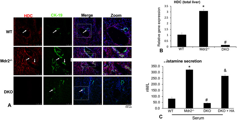
We have previously shown that Mdr2−/− mice and human PSC have increased HDC expression and histamine secretion compared to controls 1, 2; therefore, we evaluated how genetic knockdown of the HDC/histamine axis may impact liver phenotypes seen in Mdr2−/− mice. Further, our work has shown that (i) cholangiocytes regulate proliferation and damage via autocrine release of histamine 22 and (ii) HDC−/− mice have decreased global HDC expression and histamine levels 23. Therefore, to deplete the HDC/histamine axis, we crossed Mdr2−/− mice with HDC−/− mice to create a homozygous, DKO colony (see detailed methods). We first verified that HDC expression (upregulated in Mdr2−/− mice compared to WT mice) was depleted in DKO mice by immunofluorescence in tissue sections (co-stained with CK-19 to identify bile ducts) and qPCR in total liver (Figure 1A-1B). Next, we found that serum histamine levels (increased in Mdr2−/− mice) were decreased in DKO mice compared to Mdr2−/− mice (Figure 1C). Although HDC has been genetically removed from Mdr2−/− mice, there is still expected histamine secretion that is primarily a result of dietary intake of histidine in the standard chow, and this is consistent with other reports that demonstrate HDC−/− mice still have some histamine secretion 2, 23. As expected, histamine treatment in DKO mice restored histamine levels that were similar to Mdr2−/− mice. (Figure 1C).
Figure 1: Depletion of the HDC/histamine axis.
HDC expression was measured in liver sections by immunofluorescence (A) and in total liver (B) by qPCR. In Mdr2−/− mice HDC expression increased compared to WT and was found to be co-localized with CK-19 (white arrows). As expected, no HDC expression was found in the DKO mouse liver (A). By qPCR we found that the gene expression of HDC increased in Mdr2−/− mice, but was mostly ablated in the DKO mouse (B). Histamine secretion increased in serum from Mdr2−/− mice compared to WT and was ablated in DKO mice (C). Chronic treatment with histamine to DKO mice increased serum secretion of histamine (C). Data are mean ± SEM of n = 12 experiments for PCR and n = 16 experiments for EIA. *p<0.05 vs. WT; #p<0.05 vs. Mdr2−/− mice and &p<0.05 vs. DKO. Representative images x20. All data is expressed as mean ± SEM.
Liver damage is regulated by HDC/histamine signaling:
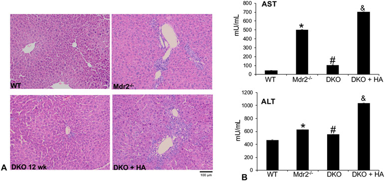
By H&E, we found that peribiliary inflammation, fibrosis, lymphocytic infiltration and necrosis, typical of the Mdr2−/− mouse model, was significantly decreased in DKO mice that lack HDC/histamine signaling. The reintroduction of histamine restored damage in DKO mice inducing similar features seen in Mdr2−/− mice, demonstrating that the HDC/histamine axis contributes to liver damage (Figure 2A). Similar findings were noted for serum AST and ALT levels showing a decrease in both markers in DKO mice that was exacerbated in DKO mice treated with histamine (Figure 2B). We also performed H&E in other organs from DKO mice and DKO mice treated with histamine and found no significant changes in morphology (data not shown).
Figure 2: Assessment of liver damage.
H&E was performed along with serum levels of ALT and AST. By H&E we found typical PSC-like damage in Mdr2−/− mice compared to WT, which were unremarkable (A). DKO mice had ameliorated liver damage including less inflammation, fibrosis, lymphocytic infiltration and necrosis; however, chronic treatment with histamine to DKO mice restored these features recapitulating Mdr2−/− mice (A). Serum AST and ALT increased in Mdr2−/− mice that was reduced in DKO mice. Histamine treatment reversed amelioration in DKO mice and AST and ALT levels were similar to Mdr2−/− mice (B). Data are mean ± SEM of n = 12 experiments for IDEXX. *p<0.05 vs. WT; #p<0.05 vs. Mdr2−/− mice and &p<0.05 vs. DKO. Representative images x20.
Genetic knockdown of HDC/histamine signaling decreases IBDM, biliary proliferation and liver inflammation:
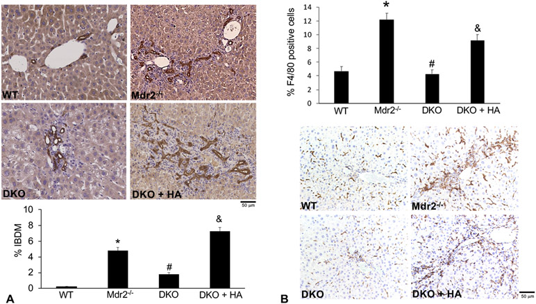
We have previously demonstrated that histamine promotes biliary proliferation in models of cholestasis 1, 2, 10, 13, 24, 25. In DKO mice, there was a significant reduction in IBDM (Figure 3A) and biliary proliferation (Supplemental Figure 1) compared to Mdr2−/− mice; however, treatment with histamine exacerbated these parameters even when compared to Mdr2−/− mice (Figure 3A and Supplemental Figure 1). These findings demonstrate that the HDC/histamine axis drives biliary damage in Mdr2−/− mice. These data are significant considering that in human PSC, characterized by increased biliary proliferation, there are increased serum histamine levels 1, 2
Figure 3: Evaluation of IBDM and inflammation.
IBDM was measured by immunohistochemistry for CK-19 (A) and inflammation by F4/80 (B). IBDM and F4/80 intensity increased in Mdr2−/− mice compared to WT, which was decreased in DKO mice (A and B). When DKO mice were treated with histamine, IBDM and F4/80 positivity increased and IBDM surpassed Mdr2−/− mice. Data are mean ± SEM of 10 representative images from n=6 mice for each group. *p<0.05 vs. WT; #p<0.05 vs. Mdr2−/− mice and &p<0.05 vs. DKO. Representative images x20.
We found that in DKO mice there was a significant decrease in Kupffer cell number shown by F4/80 staining when compared to Mdr2−/− mice. DKO mice that were treated with exogenous histamine had a significant increase in Kupffer cell number (Figure 3B). Similar trends were noted for expression of TNF-α, shown by qPCR, and for the expression of IL-6 shown by immunohistochemistry (Supplemental Figure 2A and 2B). Furthermore, we found increased serum levels of TNF-α, VEGF, FGF-β, IFN-γ, leptin, IL-1β, SCF, PDGF-BB, IL-17α, IL-4 and IL-10 in Mdr2−/− mice when compared to control; however, the expression of these factors, as well as IL-1α, G-CSF, GM-CSF, MCP-1, MIP-1α, Rantes, β-NGF, IL-2 and IL-12, was significantly decreased in DKO mice (Supplemental Figure 2C). However, DKO mice treated with exogenous histamine only had increased expression of leptin and FGF-β when compared to control treated mice (Supplemental Figure 2C).
HSC activation and hepatic fibrosis decrease in DKO mice:
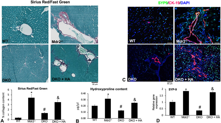
Inhibition of MC-derived histamine via cromolyn sodium or H1/H2 HR antagonist treatment reduces hepatic fibrosis and HSC activation in Mdr2−/− mice 1, 2. We evaluated hepatic fibrosis in our DKO model and found that collagen deposition shown by Sirius Red/Fast Green (Figure 4A), hydroxyproline content (Figure 4B), and the expression of pro-fibrotic genes collagen type-1a and α-SMA (Supplemental Figure 3) were decreased compared to Mdr2−/− mice. Treatment with exogenous histamine to DKO mice restored hepatic fibrosis resembling Mdr2−/− mice (Figure 4A-B and Supplemental Figure 3).
Figure 4: Hepatic fibrosis evaluation.
Hepatic fibrosis was measured by immunostaining for Sirius Red/Fast Green (A); hydroxyproline content (B) and HSC activation by immunofluorescence (C) and real-time PCR (D) for SYP-9. In Mdr2−/− mice collagen deposition and HSC activation are increased compared to WT, which is decreased in the DKO mice (A-D). When DKO mice are treated with histamine, collagen deposition/content and HSC activation are restored (A-D) and were similar or greater than Mdr2−/− mice. Data are mean ± SEM of n = 12 experiments for PCR and for staining 10 representative images from n=6 mice for each group. *p<0.05 vs. WT; #p<0.05 vs. Mdr2−/− mice and &p<0.05 vs. DKO. Representative images x20.
We noted similar changes for HSC activation demonstrated by immunofluorescence and qPCR expression for SYP-9 showing decreased activation in DKO mice compared to Mdr2−/− mice, that was restored in DKO mice treated with histamine (Figure 4C-D). Overall, these findings conclude that histamine is a key regulator of HSC activation and subsequent hepatic fibrosis in Mdr2−/− mice. However, it is still unknown if histamine directly impacts HSC activation, or mediates HSC fibrogenesis via increased neuroendocrine signaling from cholangiocytes.
Loss of the HDC/histamine axis reduces H19 signaling in DKO mice:
H19 promotes cholestatic injury and cholangiocyte proliferation via S1PR2 activation26. Similar to previous reports, we found an increase in H19 and S1PR2 expression in Mdr2−/− mice, which was significantly reduced in DKO mice (Supplemental Figure 4A, B). However, DKO mice subjected to histamine treatments had an increase in H19/S1PR2 expression when compared to control treated DKO mice (Supplemental Figure 4A, B). Furthermore, serum TBA were increased in Mdr2−/− mice when compared to WT, but reduced in DKO mice when compared to Mdr2−/− mice (Supplemental Figure 4C). However, administration of histamine to DKO mice significantly increases serum TBA levels when compared to control (Supplemental Figure 4C). S1P, a ligand for S1PR2, promotes histamine release from MCs, demonstrating a link between histamine and S1PR2 signaling27; however, these findings are the first to show potential interactions between H19 and histamine signaling.
HDC/histamine signaling targets TGF-β1 signaling via H1HR/Ca2+ signaling:
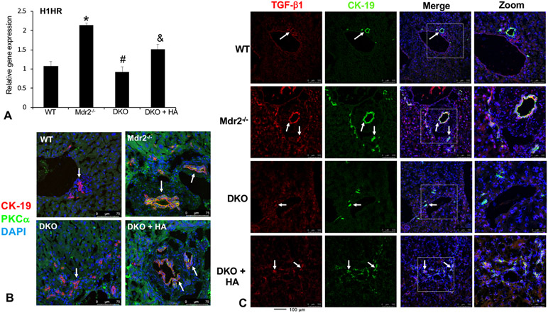
By IPA software we found that there is a relationship between histamine and TGF-β1 that can be mediated by H1HR/Ca2+ signaling (Supplemental Figure 5), which we verified in our models. Since we have previously demonstrated that H1HR can signal via the Ca2+/PKC-α pathway, we measured this in our mice. In Mdr2−/− mice we found an increase in H1HR expression (Figure 5A) and PKC-α expression (Figure 5B) compared to WT that were decreased in DKO mice. Further, when DKO mice were treated with histamine, H1HR and PKC-α expression increased (Figure 5 A-B).
Figure 5: In vivo relationship between HDC/histamine and TGF-β1 after modulation of the HDC/histamine axis.
In Mdr2−/− mice H1HR expression increases compared to WT that is lost in DKO mice (A). When DKO mice are treated with histamine, H1HR expression is restored (A). Expression of PKC-α (shown by immunofluorescence co-stained with CK-19) also increases in bile ducts from Mdr2−/− mice compared to WT, which is reduced in DKO mice (B). Again, treatment with histamine restored PKC-α expression (B). Immunofluorescence (co-stained with CK-19) shows that biliary TGF-β1 expression increases in Mdr2−/− mice, but is lost in DKO mice (C). When DKO mice are treated with histamine, biliary TGF-β1 expression increased (C). Data are mean ± SEM of n = 12 experiments for real-time PCR. *p<0.05 vs. WT; #p<0.05 vs. Mdr2−/− mice and &p<0.05 vs. DKO. Representative images x20.
By immunofluorescence, we found that TGF-β1 was upregulated in Mdr2−/− mice compared to WT. Loss of HDC/histamine signaling decreased the expression of TGF-β1 in DKO mice compared to Mdr2−/− mice (Figure 5C). This finding was further substantiated by real-time PCR and EIA which demonstrates that TGF-β1 gene expression and serum secretion of TGF-β1 significantly increased in Mdr2−/− mice that was ablated in DKO mice (Supplemental Figure 6A-B). In DKO mice treated with histamine, there was a significant increase in TGF-β1 gene expression and serum TGF-β1 level compared to Mdr2−/− mice (Supplemental Figure 6A-B).
The ablation of the HDC/histamine axis reduces VEGF signaling via downregulation of H2HR/pERK signaling:
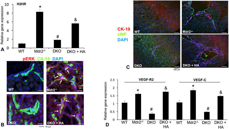
According to the IPA, there are several intermediators between histamine and VEGF including the H2HR/cAMP/pERK pathway; therefore, we evaluated these in our models. Further, our previous work has demonstrated that (i) H2HR signals primarily through Gαs coupling of cAMP/pERK 2, 10 and (ii) histamine regulates VEGF 11. By qPCR and immunofluorescence, we found that H2HR (Figure 6A) and pERK (Figure 6B) are upregulated in Mdr2−/− mice compared to WT, but are reduced in DKO mice. Further, histamine treatment in DKO mice increased these factors (Figure 6A-B). By immunofluorescence we found that vWF expression is decreased in DKO mice compared to Mdr2−/− mice and treatment with histamine restores vWF expression in DKO mice (Figure 6C). By qPCR, we found a significant increase in VEGF-R2 and VEGF-C expression in Mdr2−/− mice compared to WT, which was significantly reduced in DKO mice. Further, histamine treatment significantly increased VEGF-R2 and VEGF-C expression in DKO mice (Figure 6D).
Figure 6: Evaluation of VEGF signaling via the H2HR/ pERK pathway after modulation of HDC/histamine axis.
The expression of H2HR (A) and pERK (B) shown by qPCR and immunofluorescence, respectively increased in Mdr2−/− mice compared to WT, which was ablated in DKO mice. Treatment with histamine in DKO mice increased these parameters (A-B). The expression of vWF (C) shown by immunofluorescence increased in Mdr2−/− mice compared to WT, which was ablated in DKO mice. Treatment with histamine in DKO mice increased vWF expression (C). Both VEGF-R2 and VEGF-C expression increased in Mdr2−/− mice; however, in DKO mice, expression was lost (D). Again, treatment with histamine increased VEGF-R2 and VEGF-C expression in DKO mice. Data are mean ± SEM of n = 12 experiments for real-time PCR and n = 18 experiments for EIA. *p<0.05 vs. WT; #p<0.05 vs. Mdr2−/− mice and &p<0.05 vs. DKO. Representative images x20.
In vitro inhibition of H1HR decreases TGF-β1 signaling, whereas blocking H2HR decreases VEGF-C signaling:
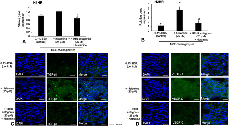
Histamine treatment increased both H1HR and H2HR gene expression, which was blocked when respective receptors were inhibited (Figure 7A). Further, the expression of TGF-β1 was increased by histamine treatment and reversed when H1HR is inhibited (Figure 7B). Finally, VEGF-C expression increases with histamine treatment which is subsequently blocked when H2HR is inhibited (Figure 7C).
Figure 7: In vitro, the HDC/histamine axis regulates TGF-β1 and VEGF signaling via H1HR and H2HR-mediated pathways, respectively.
Histamine treatment increased the expression of H1HR and H2HR that was subsequently blocked when the respective receptor was inhibited (A-B). Biliary TGF-β1 expression and VEGF-C expression increased after histamine treatment shown by immunofluorescence (C and D). Inhibition of H1HR decreased TGF-β1 expression (C), whereas blocking H2HR decreased VEGF-C expression (D). Data are mean ± SEM of n = 9 experiments for qPCR. *p<0.05 vs. basal; #p<0.05 vs. histamine treatment. Representative images x20.
DISCUSSION
PSC is characterized by increased inflammation and bile duct damage, both of which are aggravated by histamine. Our previous study has shown that blocking histamine signaling using pharmacological inhibitors (H1/H2HR blockers) or genetic models (HDC−/− mice) inhibits liver damage and fibrosis 2, 11. In the current study, we demonstrated that genetic knockdown of HDC/histamine signaling (in DKO mice) ameliorates the typical damage seen in Mdr2−/− mice including liver damage, biliary proliferation, inflammation, hepatic fibrosis, and vascular cell alterations. When DKO mice were treated chronically with histamine, damage was recapitulated and similar (or worse) to that seen in Mdr2−/− mice. Further, we found that histamine-mediated H1HR increases biliary TGF-β1 via PKC-α signaling, whereas H2HR increases cholangiocyte VEGF-C signaling via pERK activation. Taken together, these data support the concept that histamine is key to PSC progression and hepatic damage, and inhibition of this axis may be a therapeutic tool for patients.
In our previous work we found that both HDC and histamine levels are significantly increased in patients with PSC 1, 2, 13 suggesting that this axis is both a potential biomarker and also contributes to disease progression. In fact, serum histamine levels have been shown to be a potential staging marker for hepatocellular carcinoma (HCC) since patients with early stage HCC display a significant increase in histamine levels compared to controls 28. Aside from PSC, patients with biliary atresia (BA) were also found to present with significantly increased levels of hepatic histamine which positively correlated with increased hepatic fibrosis 29. In addition, both HDC and H1HR expression were upregulated in BA patient livers compared to controls 29 suggesting that these may be potential therapeutic targets for BA.
To examine the role that HDC/histamine plays during PSC progression, we generated a homozygous double knockout strain wherein this axis is depleted. Our model is supported by data showing that HDC expression and histamine secretion are significantly reduced in DKO mice compared to Mdr2−/− mice. Serum histamine levels are not completely absent since these mice also get histamine in their diet, which is consistent with our previous findings and other work from Ohtsu, et al. 11, 12, 23. In DKO mice, the degree of liver damage was significantly reduced compared to Mdr2−/− mice again suggesting that this axis promotes biliary damage and hepatic fibrosis. Previously we described the genetic knockdown of HDC on non-alcoholic fatty liver disease (NAFLD) progression and found that depletion of HDC decreases NAFLD-induced biliary damage and hepatic fibrosis; however, HDC−/− mice fed high fat diet had increased steatosis presumably due to the dysregulation of leptin/histamine signaling 12. The depletion of HDC not only affects cholangiocyte proliferation and hepatocyte ballooning, it also plays a role in critical brain function. A number of studies have demonstrated that HDC−/− mice have exaggerated features of human Tourette’s Syndrome 30, 31 thus suggesting a prominent role of histamine during proper brain function.
Although histamine is not considered a growth hormone, it has been shown to have trophic effects on cellular proliferation acting via HRs and subsequently activating secondary signaling pathways 9, 10, 32. In support of our current study, it has been demonstrated that histamine induces proliferation of both human conjunctival fibroblasts and pterygium fibroblasts 33 which was blocked by inhibition of H1HR. In a recent study we found that inhibition of either H1HR or H2HR decreases biliary damage, inflammation and hepatic fibrosis in Mdr2−/− mice 2. Further, inhibition of H1 or H2 HRs significantly reduced histamine secretion in Mdr2−/− mice 2 and our current study supports this feedback loop between HDC/histamine and HRs by demonstrating that treatment with histamine activates differential receptor signaling in DKO mice via H1HR/PKC-α and H2HR/pERK.
Reintroduction of histamine into DKO mice (that have ameliorated damage) caused a recapitulation of liver damage, biliary proliferation, inflammation and hepatic fibrosis, which are all features of DR. Causes of DR are primarily related to reactive cholangiocytes that acquire and secrete a number of neuroendocrine factors including secretin, angiogenic factors, melatonin and histamine 34, 35. Besides, PSC, DR is also a feature of autoimmune hepatitis as indicated by enhanced portal and lobular inflammation and centrilobular necrosis in biopsied patients 36. In our study we found that ablation of the HDC/histamine axis reduces DR and, as proof that histamine is a key component of DR, treatment with histamine to DKO mice restored DR similar to that of Mdr2−/− mice. The biliary tree responds to injury including increased toxic bile acid content (found in Mdr2−/− mice) by increasing the number of bile ducts to allow for increased bile flow 20, 37. However, this reparative mechanism can become damaging when biliary proliferation becomes exacerbated. Our previous work demonstrating that histamine increases biliary mass and proliferation following BDL or after CCl4 treatment suggests that increased histamine levels (from both cholangiocytes and MCs, which infiltrate after liver damage) may aggravate liver damage, specifically hepatic fibrosis 24.
H19 promotes biliary proliferation and liver injury via S1PR2 activation in murine models of cholestasis20, 26. Our findings indicate that HDC/histamine signaling promotes H19 and S1PR2 expression. Currently, no studies exist that indicate a link between H19 and histamine signaling; however, it is known that MCs can be activated by S1P (a specific ligand for S1PR2) to release histamine27, 38. Aside from S1P, the conjugated bile acid taurocholic acid (TCA) is able to activate S1PR2 signaling37. Early work found that TCA is able to induce MC activation and subsequent histamine release39; therefore, there may be a link between TCA/S1PR2 and histamine signaling. Signaling between HDC/histamine and H19/S1PR2 has yet to be elucidated, and future work should evaluate this potential interaction.
Besides histamine, other factors also contribute to DR and the progression of liver damage and hepatic fibrosis including VEGF 40, 41 and TGF-β1 34. In fact, we found that histamine interacts and activates TGF-β1 and VEGF-C levels in cholangiocytes. By both IPA and in vitro work, we found histamine treatment increases TGF-β1, as well as VEGF-C expression and, in vivo, DKO mice treated with histamine display elevated levels of both factors. Our previous study has shown that MC-derived histamine regulates TGF-β1 1, but our current study also demonstrates a link between H1HR and TGF-β1. To support our findings, during would healing, histamine increased TGF-β1 in fibroblasts, which was blocked by pre-treatment with an H1HR inhibitor 42. Our studies found that increased biliary TGF-β1 was induced by histamine treatment both in vitro and in vivo and since cholangiocytes express HDC and secrete histamine 22, we suspect that histamine treatment in DKO mice may regulate biliary TGF-β1 via H1HR/PKC-α signaling since TGF-β1 activation was reduced with H1HR inhibition.
In addition, previous data from our lab showed that downregulation of miR-125b induces increased HDC/histamine and VEGF-A and VEGF-C via HIF1-α 11. In our current study, we find that histamine regulates VEGF-C via H2HR/pERK, which are upregulated in our Mdr2−/− mice, but decreased in DKO mice. Histamine treatment to DKO mice increases these angiogenic factors and signaling pathway. Histamine and VEGF signaling have long been studied during wound healing 43, 44 and it’s been shown that specific HR signaling may also regulate VEGF response. For example, during colorectal cancer, inhibition of H2HR decreased angiogenic activity 45 and we have found that blocking either H1 or H2 HR decreases VEGF in Mdr2−/− mice and in nu/nu mice with xenograft tumors 2. The link between angiogenesis and PSC progression is not well known, therefore our studies provide important information regarding this possible feature of PSC. A very early study demonstrated that, during PSC, the duct to vessel ratio remains normal; however, the capillaries appear pushed away from ducts due to increased fibrosis and further, new smaller capillaries were found within fibrous septa suggesting that vasopenia and possible angiogenesis (during later stage PSC) are players during PSC 46.
In summary, our current study supports our previous work demonstrating that the HDC/histamine axis is a critical regulator of PSC progression. Ablation of this axis using a genetically modified mouse model shows that loss of the HDC/histamine axis ameliorates liver damage and hepatic fibrosis. Further, treatment with histamine reactivates the axis via H1 and H2 HRs, thus recapitulating PSC liver damage and hepatic fibrosis. Intermediate players including TGF-β1 and VEGF-C may be target therapies for PSC via histamine regulation including using HR blockers and/or other histamine inhibitors. Further studies are warranted to fully elucidate the role of HRs in this disease, as well as the contribution of hepatic MCs, which are the prime source of histamine. Unpublished data from our lab demonstrates that reintroduction of MCs into DKO mice also exacerbates biliary damage and hepatic fibrosis and we are currently elucidating those mechanisms (Francis and Kennedy, unpublished data 2018).
Supplementary Material
Acknowledgements:
The studies were supported by a SRCS Award to GA, VA Merit Awards (1I01BX003031, HF; 4I01BX000574, GA) from the United States Department of Veteran’s Affairs, Biomedical Laboratory Research and Development Service and NIH grant (DK108959, HF) and funds from Baylor Scott & White Institute.
Footnotes
Conflict of Interest: The authors have declared that no conflict of interest exists.
Disclosures: This material is the result of work supported by resources at the Central Texas Veterans Health Care System and Richard L. Roudebush VA Medical Center. The content is the responsibility of the author(s) alone and does not necessarily reflect the views or policies of the Department of Veterans Affairs or the United States Government.
REFERENCES
- 1.Jones H, Hargrove L, Kennedy L, Meng F, Graf-Eaton A, Owens J, et al. Inhibition of mast cell-secreted histamine decreases biliary proliferation and fibrosis in primary sclerosing cholangitis Mdr2(−/−) mice. Hepatology 2016;64:1202–1216. [DOI] [PMC free article] [PubMed] [Google Scholar]
- 2.Kennedy L, Hargrove L, Demieville J, Karstens W, Jones H, DeMorrow S, et al. Blocking H1/H2 histamine receptors inhibits damage/fibrosis in Mdr2(−/−) mice and human cholangiocarcinoma tumorigenesis. Hepatology 2018March 30. doi: 10.1002/hep.29898. [Epub ahead of print]. [DOI] [PMC free article] [PubMed] [Google Scholar]
- 3.Lee CW, Ronnekleiv-Kelly S. Autoimmune Diseases of the Biliary Tract: A Review. Surg Clin North Am 2019;99:185–201. [DOI] [PubMed] [Google Scholar]
- 4.Rajapaksha IG, Angus PW, Herath CB. Current therapies and novel approaches for biliary diseases. World J Gastrointest Pathophysiol 2019;10:1–10. [DOI] [PMC free article] [PubMed] [Google Scholar]
- 5.Lee JJ, Schindera ST, Jang HJ, Fung S, Kim TK. Cholangiocarcinoma and its mimickers in primary sclerosing cholangitis. Abdom Radiol (NY) 2017;42:2898–2908. [DOI] [PubMed] [Google Scholar]
- 6.Rizvi S, Gores GJ. Pathogenesis, diagnosis, and management of cholangiocarcinoma. Gastroenterology 2013;145:1215–1229. [DOI] [PMC free article] [PubMed] [Google Scholar]
- 7.Francis H, Meng F, Gaudio E, Alpini G. Histamine regulation of biliary proliferation. J Hepatol 2012;56:1204–1206. [DOI] [PMC free article] [PubMed] [Google Scholar]
- 8.Onori P, Gaudio E, Franchitto A, Alpini G, Francis H. Histamine regulation of hyperplastic and neoplastic cell growth in cholangiocytes. World J Gastrointest Pathophysiol 2010;1:38–49. [DOI] [PMC free article] [PubMed] [Google Scholar]
- 9.Francis H, Glaser S, DeMorrow S, Gaudio E, Ueno Y, Venter J, et al. Small mouse cholangiocytes proliferate in response to H1 histamine receptor stimulation by activation of the IP3/CaMK I/CREB pathway. Am J Physiol Cell Physiol 2008;295:C499–513. [DOI] [PMC free article] [PubMed] [Google Scholar]
- 10.Francis HL, DeMorrow S, Franchitto A, Venter JK, Mancinelli RA, White MA, et al. Histamine stimulates the proliferation of small and large cholangiocytes by activation of both IP3/Ca2+ and cAMP-dependent signaling mechanisms. Lab Invest 2012;92:282–294. [DOI] [PMC free article] [PubMed] [Google Scholar]
- 11.Graf A, Meng F, Hargrove L, Kennedy L, Han Y, Francis T, et al. Knockout of histidine decarboxylase decreases bile duct ligation-induced biliary hyperplasia via downregulation of the histidine decarboxylase/VEGF axis through PKA-ERK1/2 signaling. Am J Physiol Gastrointest Liver Physiol 2014;307:G813–823. [DOI] [PubMed] [Google Scholar]
- 12.Kennedy L, Hargrove L, Demieville J, Bailey JM, Dar W, Polireddy K, et al. Knockout of l-Histidine Decarboxylase Prevents Cholangiocyte Damage and Hepatic Fibrosis in Mice Subjected to High-Fat Diet Feeding via Disrupted Histamine/Leptin Signaling. Am J Pathol 2018;188:600–615. [DOI] [PMC free article] [PubMed] [Google Scholar]
- 13.Hargrove L, Kennedy L, Demieville J, Jones H, Meng F, DeMorrow S, et al. Bile duct ligation-induced biliary hyperplasia, hepatic injury, and fibrosis are reduced in mast cell-deficient Kit(W-sh) mice. Hepatology 2017;65:1991–2004. [DOI] [PMC free article] [PubMed] [Google Scholar]
- 14.Wu N, Meng F, Zhou T, Venter J, Giang TK, Kyritsi K, et al. The Secretin/Secretin Receptor Axis Modulates Ductular Reaction and Liver Fibrosis through Changes in Transforming Growth Factor-beta1-Mediated Biliary Senescence. Am J Pathol 2018;188:2264–2280. [DOI] [PMC free article] [PubMed] [Google Scholar]
- 15.Gaudio E, Onori P, Pannarale L, Alvaro D. Hepatic microcirculation and peribiliary plexus in experimental biliary cirrhosis: a morphological study. Gastroenterology 1996;111:1118–1124. [DOI] [PubMed] [Google Scholar]
- 16.Kyritsi K, Chen L, O’Brien A, Francis H, Hein TW, Venter J, et al. Modulation of the TPH1/MAO-A/5HT/5HTR2A/2B/2C Axis Regulates Biliary Proliferation and Liver Fibrosis During Cholestasis. Hepatology 2019. [DOI] [PMC free article] [PubMed] [Google Scholar]
- 17.Popov Y, Patsenker E, Fickert P, Trauner M, Schuppan D. Mdr2 (Abcb4)−/− mice spontaneously develop severe biliary fibrosis via massive dysregulation of pro- and antifibrogenic genes. J Hepatol 2005;43:1045–1054. [DOI] [PubMed] [Google Scholar]
- 18.Kennedy L, Francis H, Invernizzi P, Venter J, Wu N, Carbone M, et al. Secretin/secretin receptor signaling mediates biliary damage and liver fibrosis in early-stage primary biliary cholangitis. FASEB J 2019. fj201802606R. [DOI] [PMC free article] [PubMed] [Google Scholar]
- 19.Wu N, Meng F, Zhou T, Han Y, Kennedy L, Venter J, et al. Prolonged darkness reduces liver fibrosis in a mouse model of primary sclerosing cholangitis by miR-200b down-regulation. FASEB J 2017;31:4305–4324. [DOI] [PMC free article] [PubMed] [Google Scholar]
- 20.Li X, Liu R, Yang J, Sun L, Zhang L, Jiang Z, et al. The role of long noncoding RNA H19 in gender disparity of cholestatic liver injury in multidrug resistance 2 gene knockout mice. Hepatology 2017;66:869–884. [DOI] [PMC free article] [PubMed] [Google Scholar]
- 21.Ueno Y, Alpini G, Yahagi K, Kanno N, Moritoki Y, Fukushima K, et al. Evaluation of differential gene expression by microarray analysis in small and large cholangiocytes isolated from normal mice. Liver Int 2003;23:449–459. [DOI] [PubMed] [Google Scholar]
- 22.Francis H, DeMorrow S, Venter J, Onori P, White M, Gaudio E, et al. Inhibition of histidine decarboxylase ablates the autocrine tumorigenic effects of histamine in human cholangiocarcinoma. Gut 2012;61:753–764. [DOI] [PMC free article] [PubMed] [Google Scholar]
- 23.Ohtsu H, Tanaka S, Terui T, Hori Y, Makabe-Kobayashi Y, Pejler G, et al. Mice lacking histidine decarboxylase exhibit abnormal mast cells. FEBS Lett 2001;502:53–56. [DOI] [PubMed] [Google Scholar]
- 24.Johnson C, Hargrove L, Graf A, Kennedy L, Hodges K, Harris R, et al. Histamine restores biliary mass following carbon tetrachloride-induced damage in a cholestatic rat model. Dig Liver Dis 2015;47:211–217. [DOI] [PubMed] [Google Scholar]
- 25.Kennedy LL, Hargrove LA, Graf AB, Francis TC, Hodges KM, Nguyen QP, et al. Inhibition of mast cell-derived histamine secretion by cromolyn sodium treatment decreases biliary hyperplasia in cholestatic rodents. Lab Invest 2014;94:1406–1418. [DOI] [PubMed] [Google Scholar]
- 26.Xiao Y, Liu R, Li X, Gurley EC, Hylemon PB, Lu Y, et al. Long Noncoding RNA H19 Contributes to Cholangiocyte Proliferation and Cholestatic Liver Fibrosis in Biliary Atresia. Hepatology 2019;70:1658–1673. [DOI] [PMC free article] [PubMed] [Google Scholar]
- 27.Oskeritzian CA, Hait NC, Wedman P, Chumanevich A, Kolawole EM, Price MM, et al. The sphingosine-1-phosphate/sphingosine-1-phosphate receptor 2 axis regulates early airway T-cell infiltration in murine mast cell-dependent acute allergic responses. J Allergy Clin Immunol 2015;135:1008–1018 e1001. [DOI] [PMC free article] [PubMed] [Google Scholar]
- 28.Abdel-Hamid NM, Abdullah AH. Serum histamine and acetylcholine variations as new noninvasive biochemical markers in staging of experimental hepatocellular carcinoma. Clin Exp Med 2019;19:115–120. [DOI] [PubMed] [Google Scholar]
- 29.Zhou K, Xie G, Wen J, Wang J, Pan W, Zhou Y, et al. Histamine is correlated with liver fibrosis in biliary atresia. Dig Liver Dis 2016;48:921–926. [DOI] [PubMed] [Google Scholar]
- 30.Baldan LC, Williams KA, Gallezot JD, Pogorelov V, Rapanelli M, Crowley M, et al. Histidine decarboxylase deficiency causes tourette syndrome: parallel findings in humans and mice. Neuron 2014;81:77–90. [DOI] [PMC free article] [PubMed] [Google Scholar]
- 31.Pittenger C Histidine Decarboxylase Knockout Mice as a Model of the Pathophysiology of Tourette Syndrome and Related Conditions. Handb Exp Pharmacol 2017;241:189–215. [DOI] [PMC free article] [PubMed] [Google Scholar]
- 32.Francis H, Franchitto A, Ueno Y, Glaser S, DeMorrow S, Venter J, et al. H3 histamine receptor agonist inhibits biliary growth of BDL rats by downregulation of the cAMP-dependent PKA/ERK1/2/ELK-1 pathway. Lab Invest 2007;87:473–487. [DOI] [PMC free article] [PubMed] [Google Scholar]
- 33.Qin Z, Fu Q, Zhang L, Yin H, Jin X, Tang Q, et al. Proliferative Effects of Histamine on Primary Human Pterygium Fibroblasts. Mediators Inflamm 2016;2016:9862496. [DOI] [PMC free article] [PubMed] [Google Scholar]
- 34.Sato K, Marzioni M, Meng F, Francis H, Glaser S, Alpini G. Ductular Reaction in Liver Diseases: Pathological Mechanisms and Translational Significances. Hepatology 2019;69:420–430. [DOI] [PMC free article] [PubMed] [Google Scholar]
- 35.Alvaro D, Mancino MG, Glaser S, Gaudio E, Marzioni M, Francis H, et al. Proliferating cholangiocytes: a neuroendocrine compartment in the diseased liver. Gastroenterology 2007;132:415–431. [DOI] [PubMed] [Google Scholar]
- 36.Verdonk RC, Lozano MF, van den Berg AP, Gouw AS. Bile ductal injury and ductular reaction are frequent phenomena with different significance in autoimmune hepatitis. Liver Int 2016;36:1362–1369. [DOI] [PubMed] [Google Scholar]
- 37.Wang Y, Aoki H, Yang J, Peng K, Liu R, Li X, et al. The role of sphingosine 1-phosphate receptor 2 in bile-acid-induced cholangiocyte proliferation and cholestasis-induced liver injury in mice. Hepatology 2017;65:2005–2018. [DOI] [PMC free article] [PubMed] [Google Scholar]
- 38.Oskeritzian CA, Price MM, Hait NC, Kapitonov D, Falanga YT, Morales JK, et al. Essential roles of sphingosine-1-phosphate receptor 2 in human mast cell activation, anaphylaxis, and pulmonary edema. J Exp Med 2010;207:465–474. [DOI] [PMC free article] [PubMed] [Google Scholar]
- 39.Quist RG, Ton-Nu HT, Lillienau J, Hofmann AF, Barrett KE. Activation of mast cells by bile acids. Gastroenterology 1991;101:446–456. [DOI] [PubMed] [Google Scholar]
- 40.Franchitto A, Onori P, Renzi A, Carpino G, Mancinelli R, Alvaro D, et al. Expression of vascular endothelial growth factors and their receptors by hepatic progenitor cells in human liver diseases. Hepatobiliary Surg Nutr 2013;2:68–77. [DOI] [PMC free article] [PubMed] [Google Scholar]
- 41.Spirli C, Villani A, Mariotti V, Fabris L, Fiorotto R, Strazzabosco M. Posttranslational regulation of polycystin-2 protein expression as a novel mechanism of cholangiocyte reaction and repair from biliary damage. Hepatology 2015;62:1828–1839. [DOI] [PMC free article] [PubMed] [Google Scholar] [Retracted]
- 42.Wolak M, Bojanowska E, Staszewska T, Ciosek J, Juszczak M, Drobnik J. The role of histamine in the regulation of the viability, proliferation and transforming growth factor beta1 secretion of rat wound fibroblasts. Pharmacol Rep 2017;69:314–321. [DOI] [PubMed] [Google Scholar]
- 43.Niu G, Ye T, Qin L, Bourbon PM, Chang C, Zhao S, et al. Orphan nuclear receptor TR3/Nur77 improves wound healing by upregulating the expression of integrin beta4. FASEB J 2015;29:131–140. [DOI] [PMC free article] [PubMed] [Google Scholar]
- 44.Numata Y, Terui T, Okuyama R, Hirasawa N, Sugiura Y, Miyoshi I, et al. The accelerating effect of histamine on the cutaneous wound-healing process through the action of basic fibroblast growth factor. J Invest Dermatol 2006;126:1403–1409. [DOI] [PubMed] [Google Scholar]
- 45.Losurdo G, Principi M, Girardi B, Pricci M, Barone M, Ierardi E, et al. Histamine and Histaminergic Receptors in Colorectal Cancer: From Basic Science to Evidence-based Medicine. Anticancer Agents Med Chem 2018;18:15–20. [DOI] [PubMed] [Google Scholar]
- 46.Washington K, Clavien PA, Killenberg P. Peribiliary vascular plexus in primary sclerosing cholangitis and primary biliary cirrhosis. Hum Pathol 1997;28:791–795. [DOI] [PubMed] [Google Scholar]
Associated Data
This section collects any data citations, data availability statements, or supplementary materials included in this article.


