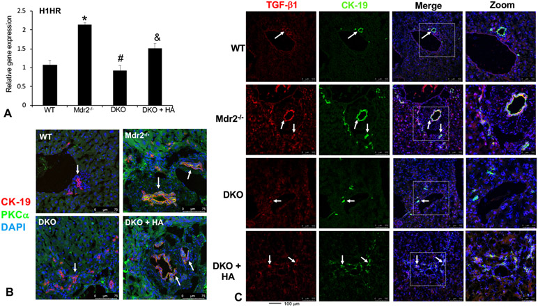
Figure 5: In vivo relationship between HDC/histamine and TGF-β1 after modulation of the HDC/histamine axis.
In Mdr2−/− mice H1HR expression increases compared to WT that is lost in DKO mice (A). When DKO mice are treated with histamine, H1HR expression is restored (A). Expression of PKC-α (shown by immunofluorescence co-stained with CK-19) also increases in bile ducts from Mdr2−/− mice compared to WT, which is reduced in DKO mice (B). Again, treatment with histamine restored PKC-α expression (B). Immunofluorescence (co-stained with CK-19) shows that biliary TGF-β1 expression increases in Mdr2−/− mice, but is lost in DKO mice (C). When DKO mice are treated with histamine, biliary TGF-β1 expression increased (C). Data are mean ± SEM of n = 12 experiments for real-time PCR. *p<0.05 vs. WT; #p<0.05 vs. Mdr2−/− mice and &p<0.05 vs. DKO. Representative images x20.

