Figure 4:

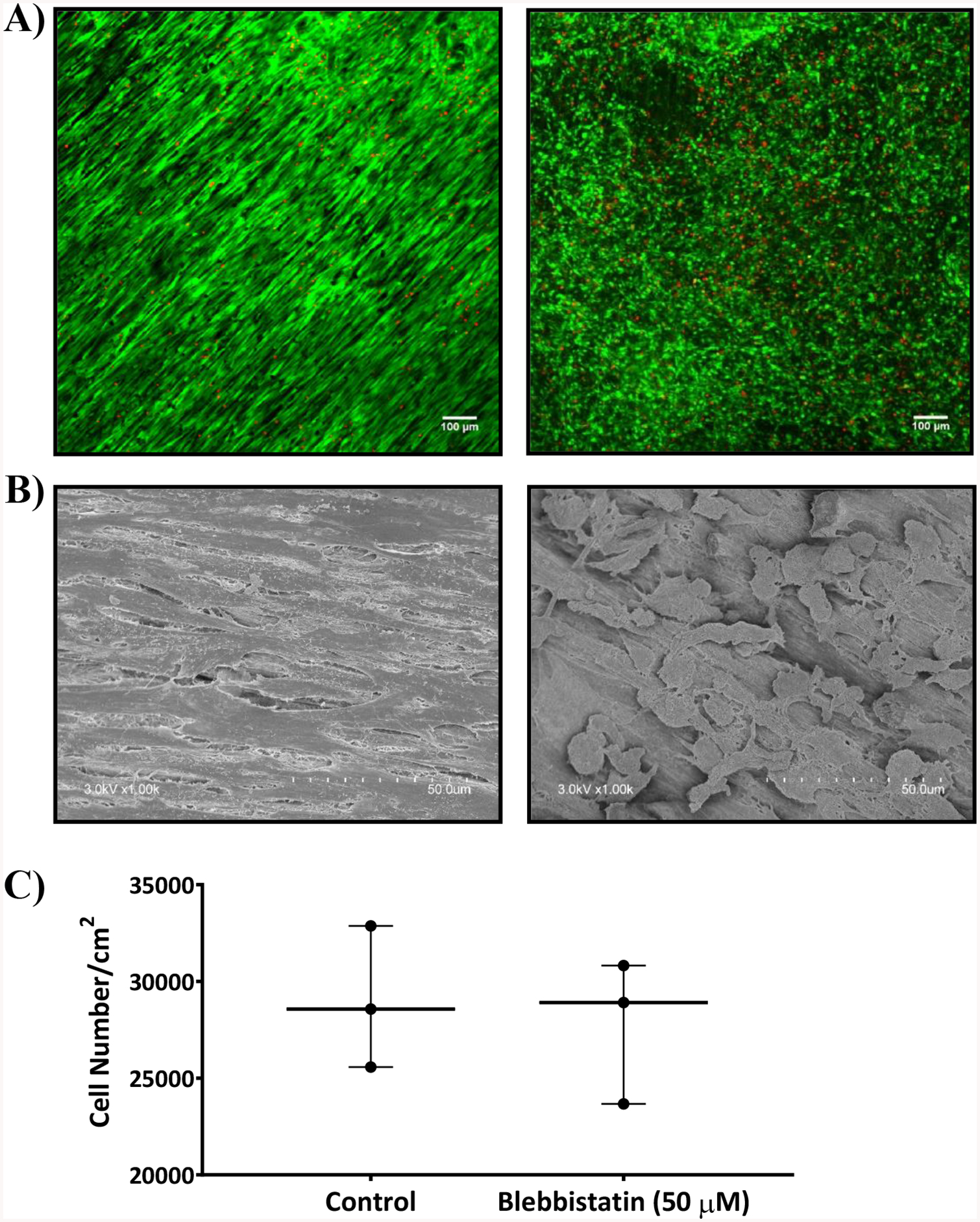
A) Confocal images B) SEM images of untreated DTS seeded with human TERT fibroblasts (left) and 50 μM blebbistatin treated DTS (right) after 6 h of seeding cells. Both were stained with calcein AM (green) and ethidium bromide (red). The cells treated with blebbistatin changed morphologically from highly branched and spindle shaped (left) to rounded (right). C) Graph showing the cell number per cm2 measured using MTS cell proliferation assay either treated or untreated with 50 μM blebbistatin. Statistical analysis was performed using unpaired two tailed t-test with n = 3.
