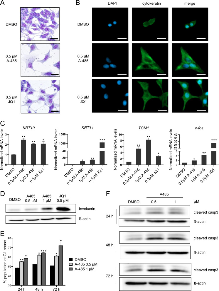
Fig. 3. A-485 induces squamous differentiation, cell cycle arrest and apoptosis.
a Hemacolor staining of HCC2429 cells incubated with 0.5 or 1 µM A-485 for 5 days. b Immunofluoresence detection of cytokeratin in HCC2429 cells incubated with 0.5 µM A-485 or JQ1 for 5 days. Scale bar = 20 µm. c Quantitative RT-PCR analysis of squamous tissue genes (KRT10, KRT14 and TGM1) and c-fos in HCC2429 cells incubated with 0.5 or 1 µM A-485 or 0.5 µM JQ1 for 5 days. Mean ± SEM from three independent experiments, ***P ≤ 0.001, **P ≤ 0.01, *P ≤ 0.05. d Immunoblot analysis of Involucrin in HCC2429 cells incubated with 0.5 or 1 µM A-485 or 0.5 µM JQ1 for 5 days. e Flow cytometry analysis of HCC2429 cells incubated with 0.5 or 1 µM A-485 for 24, 48 and 72 h. Mean ± SEM from three independent experiments, ***P ≤ 0.001, **P ≤ 0.01, *P ≤ 0.05. f Immunoblot analysis of cleaved caspase-3 in HCC2429 cells incubated with 0.5 or 1 µM A-485 for 24, 48 and 72 h.

